- Physics Lab Equipment
- Chemistry Lab Equipment
- Biology Lab Equipment
- Anatomical Lab Equipment
- Learning
- Educational Lab Equipment
- Magnets
- Agriculture
- Astronomy
- Earth Science
- Geography Lab Equipment
- Safety Lab Coat
- Laboratory
- Meteorological
- Electricity & Magnetism
- Heat
- School Lab Equipment
- Lab Glassware
- Science Lab Equipment
- Tvet Lab Equipment
- Robotics Kits Supplier and Manufacturer for Schools India
- Math Lab Equipment Manufacturers and Exporters in Ambala
- STEM Kits Manufacturers and Supplier India
- Engineering Lab Equipment
- Hospital Lab Equipment
- Water, Sanitation & Hygiene Supplies
- Cold Chain, Solar & Communication Equipment
- Nutrition
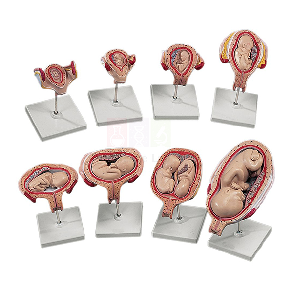
3B Human Development Set
The 3B Human Development Set (Product Code: SLE/A/134) is a series of uterus models that follows pregnancy from the early embryo to a well-formed fetus, with the embryo or fetus lifting out of each uterus for closer study. It is used in embryology, reproductive biology, midwifery and nursing teaching, and works well as a human biology model set for colleges in India. The set is supplied by Jain Scientific Equipments Pvt Ltd (ScienceLab), Ambala.
Specifications
| Parameter | Detail |
|---|---|
| Uterus models | Eight models of the uterus |
| Embryo stages | Removable embryo in the 1st, 2nd and 3rd months |
| Fetus stages | Removable fetus in the 5th and 7th months |
| 5th-month models | Four models showing twins as well as a fetus in various positions |
| Mounting | Each model mounted on a base |
Typical Use
Laid out in order on a bench, the models let students compare how the embryo grows through the first three months and how the fetus fills the uterus by the 5th and 7th months. Lifting each embryo or fetus out of its uterus makes the relationship between the developing baby and the uterine wall easy to discuss, and the 5th-month models open up topics such as twin pregnancy and fetal position that are central to obstetric and midwifery training.
Tutors often rotate small groups through the set during practical sessions, asking each group to identify the stage and describe the changes they see. Institutions equipping several teaching rooms can combine it with other pregnancy and pelvis models in a single biology model kit bulk order.
Supplier
The set reaches customers through Jain Scientific Equipments Private Limited, the Ambala, Haryana company behind the ScienceLab brand, which supplies anatomy models to schools, medical and nursing colleges and distributors. More about the company is on our company profile.
Related Human Models
Models that are commonly taught alongside this development set:
- Human Placenta Model — shows how the placenta and umbilical cord support the fetus
- Human Birth Demonstration Model — continues the story from late pregnancy to delivery through the pelvis
- Human Adult Female Pelvis Structural Model — explains the bony birth canal the fetus must pass through
- Human Embryo Development Set — an alternative gestation series for comparison
- Human Embryo Set, PVC — another uterus-and-fetus series covering the gestation period
Browse all Human Models for further reproductive and developmental anatomy aids.
Frequently Asked Questions
Which stages of pregnancy does the set cover?
It shows a removable embryo in the 1st, 2nd and 3rd months and a removable fetus in the 5th and 7th months.
What do the 5th-month models show?
Four models in the 5th month show twins as well as a fetus in various positions, which helps when teaching fetal lie and multiple pregnancy.
How many pieces will I receive?
Our listing describes eight uterus models plus the four 5th-month models, each on its own base. Please contact us to confirm the exact piece count and packing before ordering.
Can I confirm the brand and origin before buying?
Yes. The product name carries the 3B brand; ask us for brand, origin and packing details as part of your quotation.
How should the models be stored between classes?
Return each embryo or fetus to its own uterus, keep the models upright on their bases and store them away from direct sunlight and heat. Clean with a soft cloth only.
What should an institutional enquiry include?
Tell us the quantity, the delivery address, and whether you need a formal quotation or tender format. We supply buyers across India and international markets.
Request a Quote
For pricing and availability of the 3B Human Development Set, contact us or place it in the enquiry cart with any other models you need.
Last updated: 24 September 2026
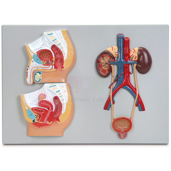
Human Male and Female Urogenital Systems Model
The Human Male and Female Urogenital Systems Model (Product Code: SLE/A/135) places a male pelvis, a female pelvis and a generalized urinary system side by side on one plaque, so students can compare how the urinary and reproductive tracts are arranged in each sex. It is designed for school biology lab equipment in India as well as nursing, paramedical and pre-medical anatomy teaching, and is made by Jain Scientific Equipments Pvt Ltd (ScienceLab), Ambala.
Specifications
| Parameter | Detail |
|---|---|
| Scale | About 3/4× life size |
| Overall size | 53 × 38 × 3 cm |
| Format | 3-part plaque mount |
| Base | Durable polymer base |
| Pelvic sections | Detailed sagittal sections of the male and female pelvis, highlighting the major urogenital structures of each |
| Urinary system | Renal artery and vein, kidneys (right side showing interior anatomy), ureters, and urinary bladder with urethra |
Typical Use
Teachers usually begin with the generalized urinary system, following blood into the kidney through the renal artery, looking at the opened right kidney, and tracing urine down the ureters to the bladder and urethra. The two sagittal pelvic sections then show where the bladder sits relative to the reproductive organs in men and women, which makes differences such as urethral length and the position of the uterus or prostate easy to point out.
Because everything is on one flat plaque, the model is convenient to prop up at the front of a class or to lay on a table for small-group revision, and it stores flat in a cupboard. It is a useful addition to any human body model kit used for reproduction, excretion and health-education topics.
Manufacturer
Manufacture is by Jain Scientific Equipments Private Limited, better known to schools and colleges by its trading name ScienceLab, and based in Ambala in the state of Haryana. You can find out more about the company on its company profile.
Related Human Models
Items that extend the same urinary and reproductive system lesson:
- Human Male Reproductive System Model Set — more detail on the male tract than the pelvic section can give
- Human Female Urogenital Model — a three-dimensional companion for the female side
- Human Kidney, Nephron, and Glomerulus Model Set — zooms into how the kidney filters blood
- Human Excretory System — a laminated wall chart to reinforce the same content
View the full Human Models category for other organ-system models.
Frequently Asked Questions
Does the model show both male and female anatomy?
Yes. It carries sagittal sections of both the male and the female pelvis, plus a generalized urinary system on the same plaque.
Can students see inside the kidney?
Yes. The right kidney is shown with its interior anatomy exposed, while the renal artery and vein, ureters and bladder with urethra complete the urinary tract.
How large is it?
The overall size is 53 × 38 × 3 cm at about 3/4× life size, so it fits on a standard classroom table.
Can it be hung on a wall?
It is a plaque mount on a polymer base. Wall-hanging fittings are not listed in our documentation, so please contact us if you plan to wall-mount it.
Is a numbered key included?
A key is not mentioned in our current product details. Ask us to confirm the teaching material supplied with the model.
What warranty applies?
Warranty terms are confirmed at the time of quotation; contact us for details.
Request a Quote
Schools, colleges and distributors in India and international markets can request pricing through our contact page or by adding this model to the enquiry cart.
Last updated: 24 September 2026
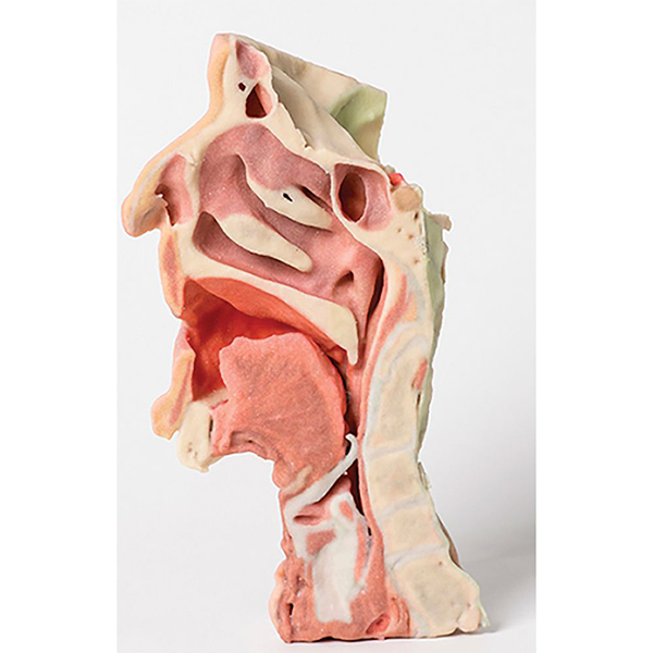
3-D Printed Head and Neck Section Model
The 3-D Printed Head and Neck Section Model (Product Code: SLE/A/136) is a midsagittally sectioned right face and neck, produced by 3-D printing to look as close as possible to a real specimen so students can study head and neck anatomy without a cadaver. Medical, dental, nursing and allied-health colleges use it for regional anatomy, and it comes from Jain Scientific Equipments Pvt Ltd (ScienceLab), Ambala, a manufacturer of human anatomical models in India.
Specifications
| Parameter | Detail |
|---|---|
| Scale | Approximately life size |
| Size | 26 × 48 × 32 cm |
| Production method | 3-D printed |
| Section shown | Midsagittal section of the right face and neck |
| Included | Laminated scientific manual |
Typical Use
A midline section is one of the most useful views in head and neck anatomy because it lets learners follow the air and food passages as a continuous route and see how deeper structures relate to the features of the face. Students can use the model to trace these pathways, identify the regions they will meet again in clinical work, and connect surface landmarks with what lies beneath them.
The laminated manual lets the model be used in self-directed study stations as well as in lectures, and because it does not need the storage conditions of a preserved specimen it can stay out in the dissection hall or skills room. It is a practical piece of lab equipment for colleges in India that want cadaver-like realism for routine teaching.
Manufacturer
The model is produced by Jain Scientific Equipments Private Limited, the Haryana company that trades as ScienceLab from Ambala and serves teaching institutions with anatomy and laboratory equipment. Read more about the company on its company profile.
Related Human Models
Other models that complement this head and neck section:
- Anatomy of the Human Neck Model — a front-on dissection of neck muscles, vessels, nerves and glands to pair with the midline view
- Head Section with Larynx Model — focuses on the upper airway and swallowing passage
- 3-D Printed Human Superficial Hand Dissection Model — another realistic 3-D printed specimen for the same teaching collection
- 3-D Printed Internal Heart Structures Model — extends 3-D printed realism to cardiac anatomy
Explore more in our Human Models range.
Frequently Asked Questions
Which side of the head does the model show?
It shows the right face and neck, sectioned along the midsagittal plane so the internal midline structures are exposed.
Why choose a 3-D printed model over a standard one?
It is made to be as realistic as possible, giving students a specimen-like appearance without the handling and storage demands of a cadaver.
What is supplied with it?
A laminated scientific manual is included to help students identify what they see.
How big is the model?
It is approximately life size and measures 26 × 48 × 32 cm.
How should it be cleaned?
Dust with a soft brush or dry cloth and avoid solvents, alcohol wipes or scrubbing, which can dull painted detail on printed surfaces.
Can you provide company documents for our purchase file?
Yes. Tell us which vendor registration details your institution needs through our contact page and we will include them with the quotation.
Request a Quote
To get a quotation for the 3-D Printed Head and Neck Section Model, contact us or add it to the enquiry cart; we supply institutions across India and international markets.
Last updated: 24 September 2026
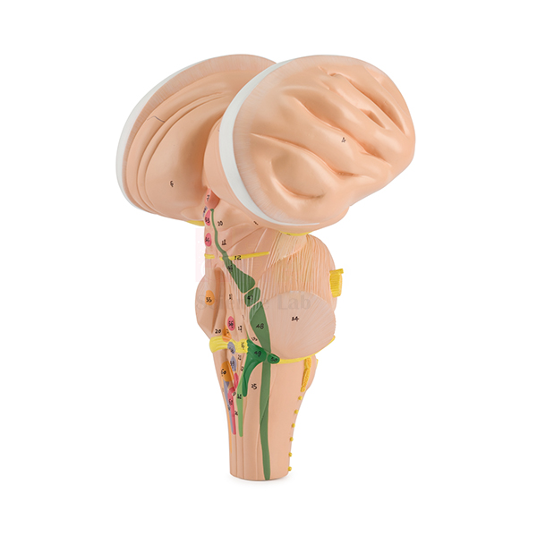
Enlarged Human Brain Stem Model
Enlarged Human Brain Stem Model (Product Code: SLE/A/137) is a 3x life-size, one-piece model of the brain stem used to teach its neurological tracts and the exiting cranial and peripheral nerves in neuroanatomy, physiotherapy, speech-language and medical courses. Measuring 26 x 17 x 13 cm, it is not on a base and comes with a numbered key to its principal structures. It is made by Jain Scientific Equipments Pvt Ltd (ScienceLab), an OEM educational laboratory equipment manufacturer, at its own factory in Ambala, India.
Specifications
| Parameter | Detail |
|---|---|
| Type | Enlarged brain stem model |
| Scale | Enlarged 3x life size |
| Size | 26 x 17 x 13 cm |
| Construction | One piece, not on a base |
| Detail shown | All the neurological tracts, plus exiting cranial and peripheral nerves |
| Labelling | Principal anatomical structures numbered; numbered key included |
| Product code | SLE/A/137 |
| Manufacturer | Jain Scientific Equipments Pvt Ltd (ScienceLab) — OEM |
| Country of origin | India (made in Ambala, Haryana) |
What Is the Enlarged Brain Stem Model Used For?
At natural size the brain stem is small and crowded, which is why cranial nerve lessons are often confusing. Enlarging it three times gives room to see each nerve leaving the stem and to follow the tracts that carry motor and sensory signals. A lecturer can hold the model up to the class, name each numbered nerve in order and link it to a quick clinical test, such as asking students to follow a finger with their eyes or to shrug their shoulders.
Being one piece without a base, it is easy to hand around a tutorial group or rest on a table at any angle. Students revising for examinations can use the numbered key to quiz themselves on the tracts and nerve exits. Colleges building a human biology model set in India often place it next to a whole-brain model to show where the stem sits before studying it enlarged.
OEM Manufacturer
ScienceLab, the brand of Jain Scientific Equipments Private Limited, is the OEM (original equipment manufacturer) of the Enlarged Human Brain Stem Model and produces it at the company's own licensed factory at Saha, Ambala, Haryana, India.
Supplying medical and allied-health colleges, dealers and importers straight from the factory means the quotation, repeat orders and any query about the numbered key come from the maker itself — just ask. More information is on the ScienceLab profile.
Related Brain Models
Models that place the brain stem in context:
- Human Brain Stem Model — another enlarged brain stem with the cranial nerve bases shown in yellow
- Enlarged Human Cerebellum Model — the neighbouring cerebellum, also shown enlarged
- Human Spinal Cord Section Model — continues the pathways downward into the spinal cord
- Human Brain Model, 15 Parts — shows where the stem sits within the whole brain
Find more in the Brain Models listing.
Frequently Asked Questions
How much is the Enlarged Human Brain Stem Model enlarged?
The Enlarged Human Brain Stem Model shows the brain stem at 3x life size and measures 26 x 17 x 13 cm, which gives room to follow each tract and exiting nerve that is crowded at natural size.
Does the Enlarged Human Brain Stem Model have a base or stand?
No. The Enlarged Human Brain Stem Model is a single piece and is not on a base, so it can be handed around a tutorial group or rested on a table at whatever angle suits the lesson.
Are the cranial nerves shown on the enlarged brain stem?
Yes. The Enlarged Human Brain Stem Model displays the exiting cranial and peripheral nerves in fine detail, along with all the neurological tracts, making it a focused aid for cranial nerve lessons.
Is a key supplied with the Enlarged Human Brain Stem Model?
Yes. A numbered key comes with the Enlarged Human Brain Stem Model and matches the numbered principal anatomical structures, so students can quiz themselves on tracts and nerve exits before examinations.
How does it differ from the Human Brain Stem Model?
Both are enlarged about three times. The Human Brain Stem Model highlights the bases of the cranial nerves in yellow, while the Enlarged Human Brain Stem Model also displays all the neurological tracts and peripheral nerves as a one-piece model without a base.
Can the Enlarged Human Brain Stem Model be taken apart?
No. The Enlarged Human Brain Stem Model is a one-piece model. For lessons that need separable parts, choose a dissectible brain such as the Human Brain Model, 15 Parts from the same range.
Who manufactures the Enlarged Human Brain Stem Model?
The Enlarged Human Brain Stem Model is manufactured by Jain Scientific Equipments Pvt Ltd (ScienceLab), the OEM, at its own licensed factory at Saha, Ambala, Haryana, India, and is supplied factory-direct to colleges, dealers and importers.
Request a Quote
For a price on the Enlarged Human Brain Stem Model, give the code SLE/A/137, quantity and delivery location in India or abroad through the contact page, or place it in the enquiry cart together with other teaching models. The manufacturer quotes directly.
Last updated: 24 September 2026
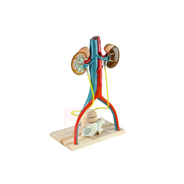
Denoyer-Geppert Free-Standing Human Urinary System Model
The Denoyer-Geppert Free-Standing Human Urinary System Model (Product Code: SLE/A/138) is a fully three-dimensional, larger-than-life replica of the male urinary apparatus and its blood supply. Unlike flat plaque models that present only half the picture, it stands on its own so students can examine the kidneys, bladder and prostate from every side. Anatomy departments, nursing schools and pharmacy colleges use it for renal and pelvic teaching, and it is supplied by Jain Scientific Equipments Pvt Ltd (ScienceLab), Ambala, a biology lab equipment supplier in India.
Specifications
| Parameter | Detail |
|---|---|
| Size | 12 × 19 × 8 in (30 × 48 × 20 cm) |
| Scale | Larger than life |
| Construction | Free-standing, fully 3-dimensional; molded entirely of non-breakable vinyl polymer |
| Part 1 | Kidneys, adrenals and blood vessels |
| Part 2 | Section of the pelvic bones with pubic symphysis, lower portion of the bladder and prostate |
| Part 3 | Upper portion of the bladder and prostate |
| Kidney section | Right kidney sagittally sectioned, revealing cortex, medulla, pyramids, calyces, pelvis and the ramifications of the renal artery and vein |
| Bladder interior | Opening the bladder exposes the folded mucosa, ureteric orifices, trigone, prostatic urethra, seminal vesicles, ejaculatory ducts and vas deferens |
| Ureters | Flexible plastic tubing |
| Key | Accompanying key identifying fifty hand-numbered features |
Typical Use
Lecturers can follow the whole urinary pathway in three dimensions: blood reaching the kidneys through the renal vessels, filtration explained on the sectioned right kidney, urine carried down the flexible ureters, and storage and outflow at the bladder. Opening the bladder brings the trigone and ureteric openings into view, while the pelvic bone section shows exactly where the bladder and prostate sit behind the pubic symphysis, a relationship that is hard to grasp from a flat diagram.
Because the model is free-standing and made of non-breakable vinyl, students can pick it up and turn it during practicals, and the key to fifty numbered features makes it suitable for spotting tests. It is a durable choice among pharmacy college lab equipment and anatomy teaching aids where renal structure underpins lessons on drug excretion and urinary disorders.
Supplier
This model is offered by Jain Scientific Equipments Private Limited, which operates under the ScienceLab name from Ambala, Haryana and sources and supplies teaching models for institutions and distributors. Our company profile gives more background.
Related Human Models
Models and charts used in the same renal and urinary teaching sequence:
- Human Kidney, Nephron, and Glomerulus Model Set — takes the lesson down to nephron and glomerulus level
- Human Kidney with Adrenal Gland Model — a closer look at a single kidney and its adrenal gland
- Human Male and Female Urogenital Systems Model — adds the female pelvis for comparison
- Male Pelvis Section Model — a sectional view of the male pelvic organs
- Human Excretory System — a laminated chart for classroom display and revision
More options are listed in our Human Models section.
Frequently Asked Questions
How is this different from a plaque-mounted urinary model?
It is fully three-dimensional and free-standing, so the urinary organs and their blood supply can be studied from all aspects rather than from one face only.
Does it show the female urinary system?
No. It represents the male urinary apparatus, including the prostate and seminal vesicles. For a male-female comparison, see the Human Male and Female Urogenital Systems Model.
How many parts does it separate into?
Three: the kidneys with adrenals and blood vessels; the pelvic bone section with the lower bladder and prostate; and the upper bladder and prostate.
Is it robust enough for student handling?
It is molded entirely of non-breakable vinyl polymer, with flexible tubing for the ureters, which suits hands-on practical sessions.
Is a key provided?
Yes. An accompanying key identifies fifty hand-numbered features on the model.
Can I confirm the brand and country of origin before ordering?
Yes. The name carries the Denoyer-Geppert brand; contact us for brand, origin and delivery details with your quotation.
Request a Quote
Send your quantity and delivery location through our contact page, or use the enquiry cart to request this model with other items. We supply India and international markets.
Last updated: 24 September 2026
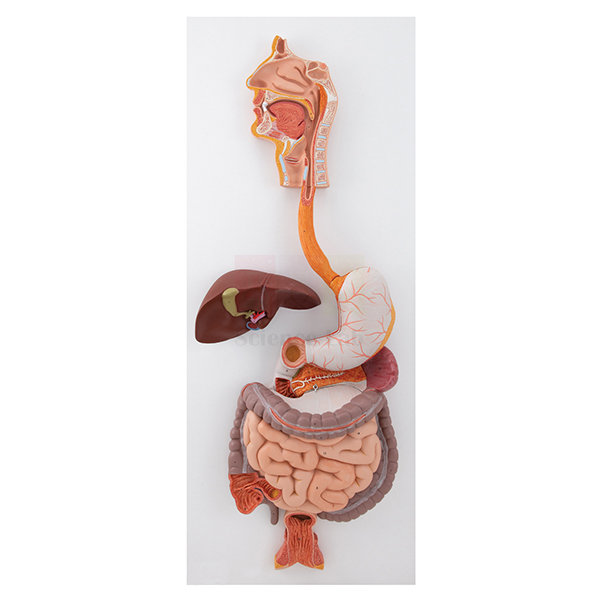
Human Digestive System Model
The Human Digestive System Model (Product Code: SLE/A/139) presents the entire digestive tract from mouth to rectum at about life size, together with the liver and pancreas, so a class can follow food through every stage of digestion on one model. A separate head section adds the chewing muscles and salivary glands. It is made by Jain Scientific Equipments Pvt Ltd (ScienceLab), Ambala, for secondary schools, colleges and nursing institutes planning a biology lab setup in India or overseas.
Specifications
| Parameter | Detail |
|---|---|
| Scale | About life size |
| Size (with base) | 92 × 32 cm |
| Second listed size | 15 × 20 cm (the part this refers to is not specified; please confirm with us) |
| Coverage | Entire digestive tract from mouth to rectum, including the liver and pancreas |
| Dissected views | Open dissection of the stomach; interior views of the small and large intestines |
| Head section | Separate section showing the musculature involved in mastication and the salivary glands |
| Key | All structures numbered and referenced on the accompanying key |
Typical Use
A digestion lesson can start with the separate head section, where students see how the chewing muscles and salivary glands begin mechanical and chemical breakdown, and then move down the tract. The opened stomach and the interior views of the small and large intestines show how the lining changes along the way, while the liver and pancreas explain where bile and digestive enzymes come from.
With every structure numbered and listed on the key, teachers can set identification exercises and practical-exam stations without extra labelling. The model suits high school biology lab equipment lists as well as nursing and nutrition courses that need a complete overview before moving on to individual organ models.
Manufacturer
Jain Scientific Equipments Private Limited makes this model and markets its educational range as ScienceLab; the company is located in Ambala, Haryana, India. Visit the company profile to learn more about us.
Related Human Models
Organ models and slides that deepen the same digestion topic:
- Human Stomach Model — a dissectible stomach for closer study of its wall and interior
- Human Liver With Gall Bladder Model — the liver lobes, gall bladder and bile ducts in detail
- Pathology of the Human Pancreas, Duodenum, and Gallbladder Model — shows common diseases of the organs seen here in healthy form
- Human Digestive Tract Model — an alternative whole-tract model on a single base
- Prepared Microscope Slide Set – Digestion (Liver, Pancreas And Tongue) — tissue sections to connect the model with microscopy
Browse the whole Human Models range for more system models.
Frequently Asked Questions
Does the model cover the whole digestive system?
Yes. It shows the entire tract from mouth to rectum, including the liver and pancreas, plus a separate head section for chewing and saliva.
Which organs are opened up?
The stomach is shown as an open dissection and the small and large intestines have interior views.
Is there a key for the numbered structures?
Yes. All structures are numbered and referenced on the accompanying key.
What are the dimensions?
The size with base is listed as 92 × 32 cm. A second size of 15 × 20 cm also appears in our records without saying which part it refers to, so please contact us to confirm before planning display space.
How should it be looked after?
Wipe with a soft, lightly damp cloth, keep it out of direct sunlight, and store the separate head section with the main model so it is not misplaced.
What information do you need for a school or college quotation?
The number of units, the delivery address and any tender or purchase-order format you must follow. We deliver across India and to international markets.
Request a Quote
Ask for pricing on the Human Digestive System Model through our contact page, or collect it with other items in the enquiry cart and send one request.
Last updated: 24 September 2026
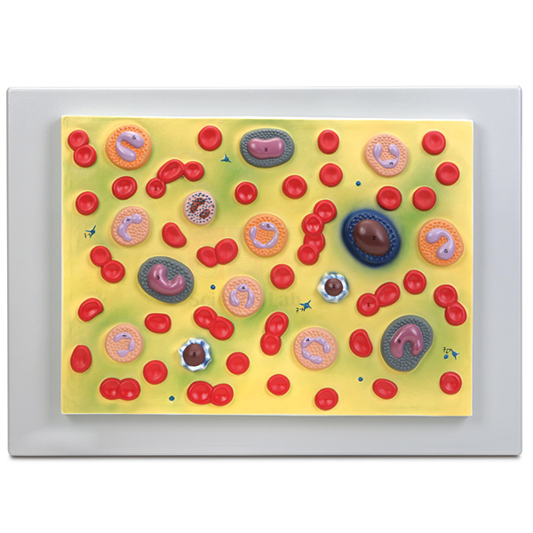
Human Blood Cell Types Model
Human Blood Cell Types Model (Product Code: SLE/A/140) is a 54 x 38 cm plaque-mounted teaching model used to show erythrocytes, platelets and 5 types of leukocytes in biology and health-science classrooms. Biology, nursing and medical laboratory technology teachers choose it because every structure is numbered on an accompanying key and each leukocyte shows its characteristic nucleus. It is made by Jain Scientific Equipments Pvt Ltd (ScienceLab), an OEM anatomical models manufacturer, at its own factory in Ambala, India.
Specifications
| Parameter | Detail |
|---|---|
| Format | Plaque mount |
| Size | 54 x 38 cm |
| Red cells | Erythrocytes |
| White cells | 5 types of leukocytes with their characteristic nuclei |
| Other formed elements | Platelets |
| Key | Structures numbered and referenced on the accompanying key |
| Product code | SLE/A/140 |
| Manufacturer | Jain Scientific Equipments Pvt Ltd (ScienceLab) — OEM |
| Country of origin | India (made in Ambala, Haryana) |
What Is the Human Blood Cell Types Model Used For?
Telling white blood cells apart is one of the first microscopy skills health-science students must master, and the shape of the nucleus is the main clue. With five leukocyte types shown side by side, a teacher can walk through the usual identification points: the lobed nucleus of the neutrophil, the two-lobed nucleus of the eosinophil, the basophil nucleus often hidden by its granules, the large round nucleus of the lymphocyte and the kidney-shaped nucleus of the monocyte. Erythrocytes, which have no nucleus, and the much smaller platelets complete the picture.
In a practical session, place the plaque where students can see it from the microscope bench. They examine a stained blood smear, sketch the cells they find and check each identification against the numbered key, which prepares them for differential leukocyte counts. Colleges building up their biology teaching aids in India often set it beside prepared slides so that theory and microscopy are covered in the same lesson.
OEM Manufacturer
Jain Scientific Equipments Private Limited, which trades as ScienceLab, is the OEM (original equipment manufacturer) of the Human Blood Cell Types Model. The plaque is made and finished at the company’s own licensed factory at Saha, Ambala, Haryana, India, rather than being sourced from a trader. The company is incorporated under CIN U29309HR2020PTC087597, and its factory at 449, HSIIDC Industrial Area, Saha, Ambala operates under Factory Licence AMB-ONLINE-CHD-J-414.
Schools, colleges, dealers and importers buy the model factory-direct, and questions about repeat orders or a replacement key go straight to the people who make it — just ask us. The company profile describes the business in more detail.
Related Laboratory Equipment
Items used with this model in a haematology practical:
- Human Blood Smear — a prepared slide for finding the same cell types under the microscope.
- Monocular Microscope 1000x, LED light — enough magnification to distinguish leukocyte nuclei on a smear.
- Haemocytometer — a counting chamber for the cell counts that follow identification.
- Blood Typing Kit, Simulated — ABO and Rh grouping practice without real blood.
The model is listed in our Animal Model category, alongside other biology teaching models.
Frequently Asked Questions
Which blood cells does the Human Blood Cell Types Model show?
The Human Blood Cell Types Model shows erythrocytes, platelets and 5 types of leukocytes on a single plaque, and each leukocyte is modelled with its characteristic nucleus so that students can compare the cells side by side before examining stained smears.
What size is the Human Blood Cell Types Model?
The Human Blood Cell Types Model is a plaque measuring 54 x 38 cm, large enough for a teacher to point out individual cells during a whole-class explanation and compact enough to store flat between practical sessions.
Does the Human Blood Cell Types Model come with a key?
Yes. Every structure on the Human Blood Cell Types Model is numbered and referenced on an accompanying key, so students can check their own identifications of red cells, platelets and each leukocyte type without waiting for the teacher.
At what magnification is the Human Blood Cell Types Model made?
The enlargement factor of the Human Blood Cell Types Model is not stated in its product details. If your course notes need it, ask us through the contact page and we will check before you order.
Who manufactures the Human Blood Cell Types Model?
Jain Scientific Equipments Pvt Ltd (ScienceLab) manufactures the Human Blood Cell Types Model as the OEM at its own licensed factory at Saha, Ambala, Haryana, India, and supplies it factory-direct to schools, colleges, dealers and importers in India and international markets.
How should the Human Blood Cell Types Model be cleaned?
Clean the Human Blood Cell Types Model by wiping the plaque gently with a soft, dry or slightly damp cloth. Avoid alcohol and solvent cleaners, which may damage the finish, and store it flat away from direct sunlight.
What does a quotation for the Human Blood Cell Types Model need?
A quotation for the Human Blood Cell Types Model needs the quantity, the delivery location and any other haematology or biology items you want on the same order; ScienceLab then quotes directly as the manufacturer.
Request a Quote
Ask for a price on the Human Blood Cell Types Model through our contact page, or collect it in the enquiry cart with your other biology items. Quotations come direct from the manufacturer.
Last updated: 24 September 2026
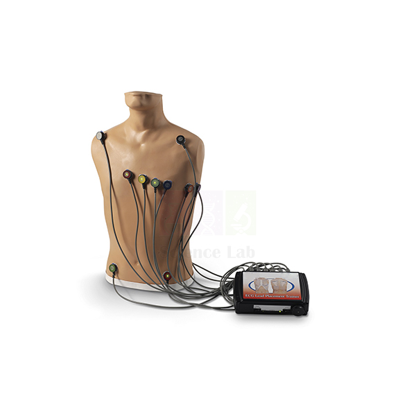
15-Lead ECG Placement Trainer
The 15-Lead ECG Placement Trainer (Product Code: SLE/A/141) is an adult trainer for learning where ECG electrodes belong and how accurately they have been placed, for recordings of up to 15 leads. Nursing, paramedical, cardiac-technology and medical students practise on its anatomical landmarks before working with patients, making it a practical aid for electrocardiograph (ECG) teaching in India lab and skills-centre settings. It is made by Jain Scientific Equipments Pvt Ltd (ScienceLab), Ambala.
Specifications
| Parameter | Detail |
|---|---|
| Type | Adult ECG electrode placement trainer |
| Lead capacity | Teaches placement for up to 15-lead ECG |
| Connection sites | 4 limb leads and V1 through V9 |
| Chest lead side | Right- or left-sided electrodes can be attached |
| Anatomical landmarks | Intercostal spaces, midclavicular line, anterior axillary line, midaxillary line and scapula |
| Signal output | Does not provide ECG output signals |
| Rhythm simulation | Rhythm and hands-free defibrillation simulations possible by adding any ECG arrhythmia simulator directly to an ECG monitor |
Typical Use
Misplaced chest electrodes are a common source of misleading ECG tracings, so this trainer focuses on the skill that comes before interpretation: finding the right spot. Learners palpate and count intercostal spaces, locate the midclavicular, anterior axillary and midaxillary lines, and then position the standard chest leads and the extra posterior and right-sided leads that make up an extended recording. The scapula landmark supports practice of the posterior positions such as V7 to V9.
Instructors can check each student’s placement against the connection sites and repeat the exercise as often as needed without a volunteer. When rhythm recognition or defibrillation drills are required, an ECG arrhythmia simulator can be connected directly to the ECG monitor, keeping this unit focused on placement. It fits well in vocational training lab equipment for paramedical and emergency-care courses.
Manufacturer
This trainer is a product of Jain Scientific Equipments Private Limited, an Ambala, Haryana manufacturer that trades as ScienceLab and supplies teaching equipment to medical, nursing and technical institutions. See the company profile for more information.
Related Human Models
Equipment often set up in the same clinical-skills room:
- Advanced Adult CPR Training Manikin — for chest-compression and rescue-breathing practice alongside ECG skills
- Human Heart Model — explains the heart anatomy that each lead views
- Stethoscope — for pairing heart-sound auscultation with electrode placement practice
- Blood Pressure Monitor — completes a basic vital-signs training station
Find more trainers and models in our Human Models category.
Frequently Asked Questions
Does the trainer produce an ECG trace?
No. It does not provide ECG output signals. Rhythm and hands-free defibrillation simulations can be run by adding an ECG arrhythmia simulator directly to an ECG monitor.
Is an arrhythmia simulator or ECG monitor included?
Neither is listed as part of this trainer. Contact us if you need advice on what to pair with it.
Which electrode positions can be practised?
There are connection sites for 4 limb leads and V1 through V9, and electrodes can be attached on the right or left side.
Is it an adult or a paediatric trainer?
It is an adult trainer, with landmarks such as the intercostal spaces, midclavicular line, axillary lines and scapula.
Are spare parts and after-sales support available?
Please ask us about spares and warranty terms when you request a quotation, as these are confirmed per order.
Can you supply several trainers for a nursing or paramedical college?
Yes. We supply institutions in India and international markets; include the quantity and delivery location in your enquiry.
Request a Quote
For a quotation on the 15-Lead ECG Placement Trainer, contact us or add it to the enquiry cart with any other skills-lab items you need.
Last updated: 24 September 2026