- Physics Lab Equipment
- Chemistry Lab Equipment
- Biology Lab Equipment
- Anatomical Lab Equipment
- Learning
- Educational Lab Equipment
- Magnets
- Agriculture
- Astronomy
- Earth Science
- Geography Lab Equipment
- Safety Lab Coat
- Laboratory
- Meteorological
- Electricity & Magnetism
- Heat
- School Lab Equipment
- Lab Glassware
- Science Lab Equipment
- Tvet Lab Equipment
- Robotics Kits Supplier and Manufacturer for Schools India
- Math Lab Equipment Manufacturers and Exporters in Ambala
- STEM Kits Manufacturers and Supplier India
- Engineering Lab Equipment
- Hospital Lab Equipment
- Water, Sanitation & Hygiene Supplies
- Cold Chain, Solar & Communication Equipment
- Nutrition
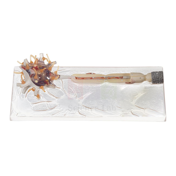
Dissectible Human Neuron
The Dissectible Human Neuron (Product Code: SLE/A/150) is a schematic model of a nerve cell and its axon, showing the medullary (myelin) sheath and Schwann’s sheath, that separates into 3 parts and stands on its own base. Its simplified, uncluttered design suits secondary school and pre-university classes that need to learn the basic parts of a neuron before moving to more detailed cell biology. It is one of the biology teaching aids in India made by Jain Scientific Equipments Pvt Ltd (ScienceLab), Ambala.
Specifications
| Parameter | Detail |
|---|---|
| Representation | Schematic nerve cell and axon |
| Sheaths shown | Medullary sheath and Schwann’s sheath |
| Dissection | Dissectible into 3 parts |
| Mounting | On stand with base |
| Size | 53 × 17 × 22 cm |
Typical Use
A schematic neuron strips away microscopic clutter so beginners can concentrate on the essentials: the cell body, the long axon that carries the impulse, and the two coverings around it. Teachers can separate the 3 parts to show how the axon sits inside its medullary sheath and how Schwann’s sheath lies outside, then reassemble the model on its stand to summarise the whole cell.
It is a practical piece of science lab equipment for class 12 and junior college biology chapters on neural control and coordination, and also works for introductory nursing and psychology courses. Because it is mounted on a stand, it can be left on display at the front of the class throughout a unit on the nervous system.
Manufacturer
The neuron is made in India by Jain Scientific Equipments Private Limited, an Ambala, Haryana enterprise that trades as ScienceLab and supplies schools and colleges with teaching models. For more on the company, open the company profile.
Related Human Models
Useful next steps after this introductory neuron:
- Neuron Model — a highly magnified neuron with organelles for advanced classes
- Human Nerve Ending and Synapse Model — shows how one neuron passes its signal to the next
- Human Nervous System Model — places neurons in the context of the brain, cord and spinal nerves
- Human Spinal Cord Section Model — where nerve cell bodies and fibres meet in the cord
Browse more in the Human Models category.
Frequently Asked Questions
What does “schematic” mean for this model?
It is a simplified representation of a nerve cell and axon, designed to show the main parts and sheaths clearly rather than every microscopic detail.
Which coverings of the axon are shown?
The medullary sheath and Schwann’s sheath.
Into how many parts does it separate?
Three. When assembled, it stands on its stand with base for display.
Is it suitable for school students?
Yes. The schematic style matches secondary and pre-university syllabi; for organelle-level detail, see the Neuron Model.
How big is it?
It measures 53 × 17 × 22 cm.
What should a school include in its purchase enquiry?
The number of models, the delivery address and any quotation format required by your school or department. We supply across India and to international markets.
Request a Quote
To receive a quotation for the Dissectible Human Neuron, contact us or add it to the enquiry cart.
Last updated: 24 September 2026
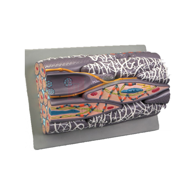
Microstructure of Smooth Muscle Model
The Microstructure of Smooth Muscle Model (Product Code: SLE/A/151) is a block model of a greatly enlarged bundle of smooth muscle fibers, revealing the structures inside and between the cells, from the nucleus, mitochondria and sarcoplasmic reticulum to the dense bodies and contractile myofilaments that produce contraction. Histology, physiology and zoology lab equipment buyers in India choose it to make an invisible tissue visible to a whole class. It is made by Jain Scientific Equipments Pvt Ltd (ScienceLab), Ambala.
Specifications
| Parameter | Detail |
|---|---|
| Type | Block model of a greatly enlarged bundle of smooth muscle fibers |
| Size | 28 × 16 × 11 cm |
| Base | Mounted on a durable polymer base |
| Structures shown | Significant extra- and intracellular structures, including sarcoplasmic reticulum, nucleus, mitochondria, dense bodies and contractile myofilaments |
Typical Use
Smooth muscle lines the walls of hollow organs and blood vessels, and its contraction works differently from the striated muscle students already know. On this block model, the nucleus, the mitochondria and the dense bodies that anchor the contractile myofilaments give the teacher a clear basis to explain why smooth muscle has no visible striations and how it can contract slowly and steadily.
Lecturers typically use it alongside prepared microscope slides of muscle tissue, so students first see the enlarged three-dimensional structure and then recognise the same tissue under the microscope. It suits undergraduate zoology, physiology, pharmacy and medical histology practicals.
Manufacturer
The model is manufactured by Jain Scientific Equipments Private Limited, an Indian company in Ambala, Haryana; its products for schools, colleges and laboratories are sold under the name ScienceLab. Details are on our company profile.
Related Human Models
Items for a complete muscle-tissue lesson:
- Skeletal Muscle Fiber Model — the striated counterpart, for a side-by-side comparison
- Microstructure of Artery and Vein Model — shows where smooth muscle layers sit in vessel walls
- Microslide Animal Tissue Set 2 Muscle, Bone, Connective — photographed microscope views of muscle and related tissues
- Micro Slide Viewer — for viewing the Microslide image strips without a microscope
For other microstructure models, see the Human Models range.
Frequently Asked Questions
Which cell structures can students identify?
The sarcoplasmic reticulum, nucleus, mitochondria, dense bodies and contractile myofilaments, plus significant structures between the fibers.
What magnification is the model?
It is described as greatly enlarged; an exact magnification is not stated in our documentation. Contact us if you need this figure.
How does it differ from a skeletal muscle model?
Smooth muscle has no sarcomere striations; its myofilaments are anchored to dense bodies, which this model shows clearly. Pair it with the Skeletal Muscle Fiber Model for comparison.
Where is smooth muscle found in the body?
In the walls of the digestive tract, blood vessels, airways, bladder and uterus, where it works involuntarily.
What size is it?
The block measures 28 × 16 × 11 cm and sits on a durable polymer base.
Can overseas universities order it?
Yes. We supply institutions and distributors in India and international markets; send the quantity and destination for a quotation.
Request a Quote
Ask for a quotation via our contact page, or place the Microstructure of Smooth Muscle Model in the enquiry cart with other histology items.
Last updated: 24 September 2026
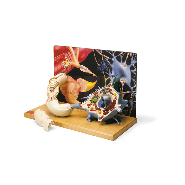
Denoyer-Geppert 3-D Human Motor Neuron Model
The Denoyer-Geppert 3-D Human Motor Neuron Model (Product Code: SLE/A/152) is a diorama that reproduces a motor nerve cell at more than 2,500 times life size against a colour backdrop of associated neurons, and follows its axon all the way to the neuromuscular junction on a skeletal muscle. Physiology, neuroscience, pharmacology and sports-science departments use it to connect nerve-cell structure with how muscles are made to contract. It is supplied by Jain Scientific Equipments Pvt Ltd (ScienceLab), Ambala, a biology lab instruments supplier.
Specifications
| Parameter | Detail |
|---|---|
| Magnification | More than 2,500× life size |
| Size | 43 × 20 × 28 cm |
| Base | Mounted on a hardwood base |
| Format | Diorama: detailed motor nerve cell with a colour backdrop showing a network of associated neurons |
| Removable section | A section of the axon lifts off to show the layers of the myelin sheath, the neurolemma and the Schwann cell that formed them |
| Neuromuscular detail | Convergence of multiple axons into a motor nerve fiber, ending at the neuromuscular junction of a skeletal muscle, with synaptic vesicles carrying neurotransmitters |
| Other highlights | Cellular organelles, branching dendrites, communicating synapses, and a myelin-wrapped axon with node of Ranvier |
| Key | Illustrated key with 54 identified features |
Typical Use
Most neuron models stop at the cell itself; this diorama carries the story through to its effect. Students can start with the dendrites and synapses receiving input, look at the organelles in the cell body, follow the myelinated axon past a node of Ranvier, and then see several axons converge into a motor nerve fiber that ends on skeletal muscle. Lifting away the axon section shows how the Schwann cell wraps the myelin layers and neurolemma around the fibre.
The synaptic vesicles at the neuromuscular junction make the model particularly useful in pharmacology lab equipment sets, where lecturers explain how neuromuscular blockers and related drugs act on transmission. With the 54-feature illustrated key, it also serves well for identification exercises in physiology practicals.
Supplier
This diorama is supplied through Jain Scientific Equipments Private Limited, the Ambala, Haryana company known to its customers as ScienceLab, which provides teaching models and lab apparatus to universities, colleges and distributors. Visit the company profile for background on the company.
Related Human Models
Complementary models and media for teaching nerve-to-muscle signalling:
- Skeletal Muscle Fiber Model — the muscle fibre that the motor neuron stimulates, at the microstructure level
- Human Nerve Ending and Synapse Model — a closer look at a single synapse
- Neuron Model — a general myelinated neuron for comparison with the motor neuron
- Microslide Nerve & Muscle Action — microscope images of nerve and muscle to match the model
- Micro Slide Viewer — the viewer needed for Microslide image sets
Explore further models in our Human Models category.
Frequently Asked Questions
What makes this a diorama model?
The motor neuron is reproduced in three dimensions in front of a colour backdrop that shows a network of associated neurons, so the cell is seen in context.
Does it show the neuromuscular junction?
Yes. Multiple axons converge into a motor nerve fiber that ends at the neuromuscular junction of a skeletal muscle, complete with synaptic vesicles carrying neurotransmitters.
Can the myelin sheath be examined in layers?
Yes. A section of the axon lifts off to show the layers of the myelin sheath, the neurolemma and the Schwann cell.
How are the features identified?
An illustrated key identifies 54 features on the model.
What are the size and base?
It measures 43 × 20 × 28 cm and is mounted on a hardwood base.
Can I confirm the brand and origin before ordering?
Yes. The name carries the Denoyer-Geppert brand; ask us for brand, origin and delivery details through our contact page.
Request a Quote
Universities and distributors in India and international markets can contact us for pricing or add this model to the enquiry cart for a combined quotation.
Last updated: 24 September 2026
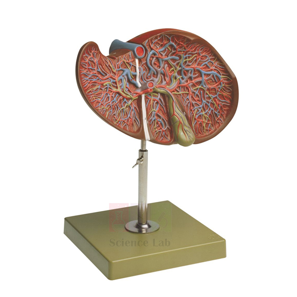
Human Liver and Gallbladder Model
Human Liver and Gallbladder Model (Product Code: SLE/A/153) is a one-part, dissected model enlarged to 1-1/2x life size, showing the internal anatomy of the human liver together with its gallbladder. The enlargement makes internal detail easier to point out to a group, and a numbered key is included. It is manufactured by Jain Scientific Equipments Pvt Ltd (ScienceLab), Ambala, for anatomy and physiology teaching in schools, nursing colleges and medical institutions, and is a useful piece among biology teaching aids in India for the digestive system.
Specifications
| Parameter | Detail |
|---|---|
| Scale | 1-1/2x life size, enlarged |
| Size | 19 x 26 x 29 cm |
| Construction | One-part, dissected model |
| Anatomy shown | Internal anatomy of the human liver with gallbladder |
| Key | Numbered key included |
Typical Use
The liver is large and solid, so its interior is hard to picture from outside views alone. Because this model is already dissected, the internal anatomy is visible the moment it is placed on the bench, with no parts to remove. Teachers use it to explain the liver’s role in producing bile, the gallbladder’s role in storing it, and how both connect to digestion of fats. Students then work through the numbered key to name each marked structure.
The one-part build suits frequent handling by younger classes, and the enlarged scale helps at the back of a lecture room. Pairing it with a liver pathology model turns a single lesson into a normal-versus-disease comparison. Clean it with a soft dry cloth and keep it away from heat sources.
Manufacturer
Manufactured by Jain Scientific Equipments Private Limited, which trades as ScienceLab from Ambala, Haryana, and produces anatomy models and teaching apparatus for institutions in India and international markets. Details about the company are on our company profile.
Related Digestive System Models
Models that fit into the same liver and biliary lessons:
- Human Liver Pathology Model — life-size liver and gallbladder showing common pathologies, for comparison
- Human Liver With Gall Bladder Model — an alternative liver and gall bladder model
- Rear Organs of the Upper Abdomen — shows the opened bile ducts, duodenum and pancreas that connect to the gallbladder
- Gallstone Model — covers gallstones and gallbladder inflammation in the biliary system
More models are listed in our Digestive System category.
Frequently Asked Questions
Why is the model enlarged?
At 1-1/2x life size, the internal features of the liver and gallbladder are larger and easier to identify during group teaching.
Does it separate into parts?
No. It is a one-part model that is already dissected to show the internal anatomy.
What size is it?
It measures 19 x 26 x 29 cm.
Is there a key?
Yes, a numbered key comes with the model.
Does it show liver disease?
No, it shows normal internal anatomy. For disease teaching, see the Human Liver Pathology Model listed above.
What about warranty and delivery time?
These are confirmed in each quotation rather than fixed on this page. Contact us with your requirement.
Request a Quote
Ask for a price on the Human Liver and Gallbladder Model through our contact page, or add it to the enquiry cart if you are ordering several anatomy models together.
Last updated: 24 September 2026
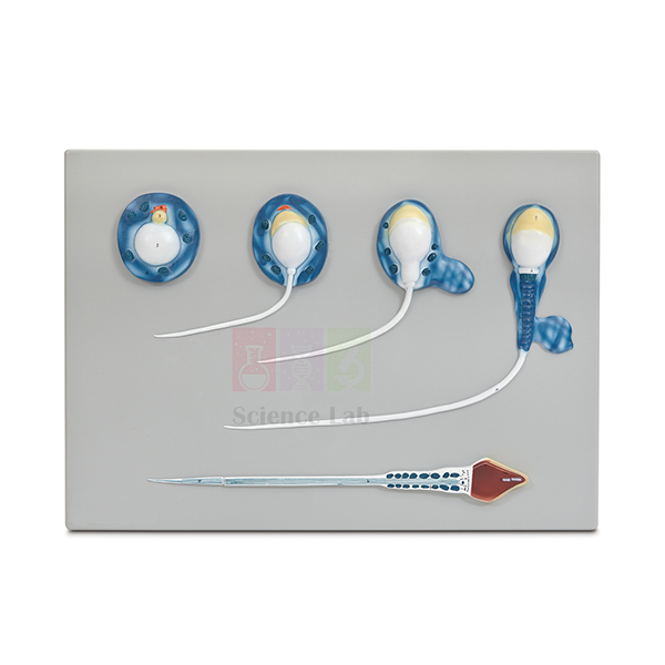
Spermatogenesis Model
The Spermatogenesis Model (Product Code: SLE/A/154) is an enlarged teaching model that follows the final phase of sperm formation, from the spermatid to the mature spermatozoon. Magnified about 10,000 times, it turns a microscopic change in cell shape into something a whole class can see from their seats, making it a practical choice among biology teaching aids for reproduction and cell biology units in senior secondary schools, colleges and nursing or paramedical institutes. It is manufactured by Jain Scientific Equipments Pvt Ltd (ScienceLab), Ambala, India.
Specifications
| Parameter | Detail |
|---|---|
| Process shown | Spermatogenesis from the spermatid to the mature spermatozoon |
| Magnification | Enlarged about 10,000x |
| Size | 53 x 38 x 5 cm |
| Mounting | Durable polymer base |
| Identification | Supplied with key |
Typical Use
In lessons on gametogenesis, the model gives students a fixed reference while the teacher explains how a rounded spermatid is remodelled into a streamlined sperm cell: the nucleus becomes compact, a cap forms over the head and a tail develops for movement. Because the whole sequence is presented on one mounted model, learners compare changes in shape and size directly instead of piecing them together from separate textbook drawings, and the key lets them check their own identifications during revision.
For a stronger practical link, pair the model with a prepared slide of mammalian testis: students first locate developing cells in real tissue under the microscope, then return to the enlarged model to see what those cells become. Used this way, it sits naturally within a human biology model set built around the reproductive system.
Manufacturer
ScienceLab is the brand name under which Jain Scientific Equipments Private Limited, a company based in Ambala, Haryana, makes anatomical models and laboratory apparatus for teaching institutions. The company profile describes its background and product range in more detail.
Related Laboratory Equipment
Items that help teach the same topic, from tissue level to the finished gamete:
- Ts Of Testis (Mammalian) — prepared slide of the tissue in which sperm are produced
- Mitosis & Meiosis Set, Microscope Slides — covers the cell divisions that come before the spermatid stage
- Human Sperm Model — a base-mounted model of the finished male gamete for introductory lessons
- Human Male Reproductive System Model Set — places sperm production in its organ-level context
See the complete Human Models range for other reproduction and anatomy teaching models.
Frequently Asked Questions
Which part of sperm development does this model show?
It shows spermatogenesis from the spermatid to the mature spermatozoon. If you need earlier stages such as spermatogonia or spermatocytes represented, please contact us before ordering, as these are not listed in the product documentation.
What are the dimensions?
The model measures 53 x 38 x 5 cm and is mounted on a durable polymer base.
How do students identify the stages?
A key is supplied with the model, so each stage can be matched to its name during lessons or self-study.
Why is the model enlarged about 10,000 times?
Sperm cells and the cells they develop from are microscopic; the enlargement makes their changes visible to a full class without a microscope. It complements slide work rather than replacing it.
How should the model be cleaned?
Wipe it with a soft, slightly damp cloth and let it dry; avoid solvents and abrasive cleaners, which can damage the finish. Store it away from direct sunlight.
Is a warranty offered?
Warranty terms are not stated in the product documentation, so please ask us for the terms that apply to your order.
Can you supply several units or ship abroad?
Yes. We supply schools, colleges and distributors in India and international markets; send the quantity and delivery location through our contact page for a quotation.
Request a Quote
To request pricing or a formal quotation for the Spermatogenesis Model, contact our sales team or add the item to your enquiry cart together with any related models you need.
Last updated: 24 September 2026
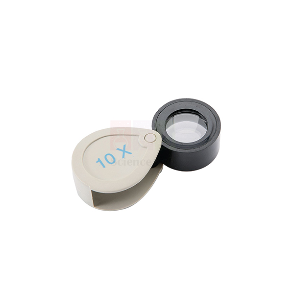
Pocket Magnifier
Pocket Magnifier (Product Code: SLE/M/86) is a folding 10X magnifier loupe used to examine leaves, insects, rocks, fabrics and fine print during field trips, nature walks and lab practicals. Its two plano-convex lenses sit in a mount that folds into the case, so the glass stays protected in a pocket or kit bag, and the whole unit is lightweight. It is supplied by Jain Scientific Equipments Pvt Ltd (ScienceLab), Ambala, India.
Specifications
| Parameter | Detail |
|---|---|
| Magnification | 10X |
| Optics | Two plano-convex lenses |
| Design | Lens mount folds into the case |
| Weight | Lightweight |
| Dimensions | 15 × 25 mm |
| Product code | SLE/M/86 |
| Supplied by | Jain Scientific Equipments Pvt Ltd (ScienceLab), Ambala, India |
What Is the Pocket Magnifier Used For?
A 10X pocket lens is the first optical tool most students carry into the field. In botany it shows leaf hairs, vein networks and the parts of small flowers; in zoology it reveals insect wing veins, spider eyes and the scales on a moth wing; in geography and geology it helps judge grain size and spot crystals in a rock sample. Teachers also use it for checking fabric weave, seed surfaces and printed detail in general science lessons.
Pairing two plano-convex lenses gives a stronger magnification in a small package than a single simple lens. For a sharp view, hold the Pocket Magnifier close to the eye and bring the specimen towards it until it snaps into focus, working in good daylight or under a lamp. Fold the lens back into its case after use to keep it free of scratches and grit. It is a simple way to give every child a hand lens magnifier in school science lessons across India.
Supplier
Jain Scientific Equipments Private Limited, the company that trades as ScienceLab in Ambala, Haryana, supplies the Pocket Magnifier to schools, colleges, nature clubs and dealers. We source it together with other magnifiers and microscopes, so a class set of field lenses can be added to a larger laboratory order. Visit our profile page to learn more about the company.
Related Microscope Products
Other magnifiers and viewers for the same observation lessons:
- Magnifier, Foldable — another folding magnifier style to compare.
- Magnifying Glass — a larger hand-held lens for group viewing.
- Micro Slide Viewer — 50x viewing of prepared specimens without power.
- Stereomicroscopes — for closer study of specimens collected in the field.
- Microfiber Lens Cloth — to wipe fingerprints from the lenses.
More optical items are listed in our Microscope category.
Frequently Asked Questions
What magnification does the Pocket Magnifier give?
The Pocket Magnifier gives 10X magnification from two plano-convex lenses used together. That is enough to see fine hairs on leaves, insect wing veins and mineral grains that are hard to make out with the naked eye.
How are the lenses of the Pocket Magnifier protected?
The lens mount of the Pocket Magnifier folds into its case. When closed, the lenses are shielded, so the magnifier can travel in a pocket, pencil box or field bag without the glass being scratched or picking up grit.
What size is the Pocket Magnifier?
The Pocket Magnifier’s listed dimensions are 15 × 25 mm, and it is lightweight enough to carry in a shirt pocket. If your tender needs the closed case size or the weight, please ask us through the contact page.
How should students use a 10X pocket magnifier?
Hold the Pocket Magnifier close to the eye and move the specimen, not the lens, until it comes into sharp focus. Good light from a window or lamp makes a big difference at 10X, especially with dark specimens.
Who supplies the Pocket Magnifier?
Jain Scientific Equipments Pvt Ltd (ScienceLab), Ambala, India, supplies the Pocket Magnifier in class quantities to schools, colleges, nature clubs and distributors in India and international markets, often alongside microscopes and slide accessories.
What details are needed to quote the Pocket Magnifier?
For a Pocket Magnifier quotation, mention product code SLE/M/86, the number of pieces, the delivery address and any other biology items on your list. Submit these through the contact page or the enquiry cart.
Request a Quote
Ask for a price on the Pocket Magnifier (SLE/M/86) in the quantity you need through our contact page, or add it to the enquiry cart with other field and lab items.
Last updated: 24 September 2026
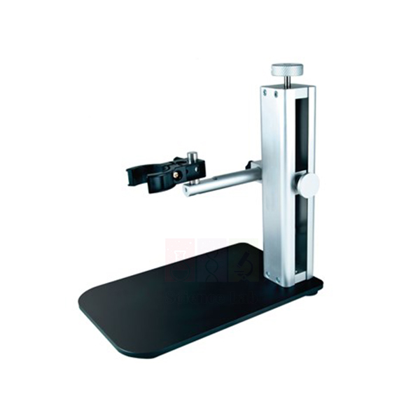
Microscope Stand
Microscope Stand (Product Code: SLE/M/87) is a precision focusing stand used to hold a compact microscope steady above a specimen for sharp viewing and imaging in classrooms, inspection benches and research labs. Built from stainless steel and lightweight aluminium, it combines a fine-focus knob, quick-release vertical movement, an adjustable safety stop and a 15 cm swivelling arm. It is supplied by Jain Scientific Equipments Pvt Ltd (ScienceLab), Ambala, India, among our laboratory instruments.
Specifications
| Parameter | Detail |
|---|---|
| Size | W 15 × L 22 × H 23 cm |
| Construction | Resilient stainless steel and lightweight aluminium |
| Focusing | Precise fine-focus adjustment knob, useful at high magnification |
| Vertical movement | Quick-release button for fast, convenient positioning |
| Safety stop | Adjustable; prevents the microscope from touching the specimen |
| Horizontal arm | Adjustable, 15 cm range, 360-degree rotation |
| Additional arm | Included; increases effective range for larger objects |
| Compatibility | Listed as compatible with all D models; confirm your microscope model with us |
| Product code | SLE/M/87 |
| Supplied by | Jain Scientific Equipments Pvt Ltd (ScienceLab), Ambala, India |
What Is the Microscope Stand Used For?
Compact and hand-held microscopes are convenient, but at higher magnification the slightest hand tremor blurs the picture and makes photographs impossible. Clamping the microscope into this stand keeps it rock-steady, so students can study coins, circuit boards, textiles, insects or rock surfaces and capture clear images for reports or projection.
A good routine is to lower the microscope with the quick-release button until it is roughly in position, set the safety stop just above the specimen height, and then finish with the fine-focus knob. The stop protects both the lens and delicate samples from contact. Swing the 15 cm arm through its 360-degree rotation to reach different parts of a board or specimen tray, and fit the additional arm when the object is too large to sit under the standard reach. It makes a practical addition to optics lab equipment in India’s school and college labs.
What to Specify When Ordering
- The make and model of the microscope you plan to mount, so fit can be confirmed.
- The size of the largest objects you intend to view.
- Number of stands required.
Supplier
The Microscope Stand is sourced and supplied by Jain Scientific Equipments Private Limited, whose trading name is ScienceLab, from Ambala, Haryana. We offer it to schools, colleges, research labs and dealers, and it can be quoted with digital microscopes and lighting so a complete viewing station arrives on one order. For vendor registration and export paperwork, the supplying company holds IEC AAECJ8618A and GSTIN 06AAECJ8618A1ZT. Read more on our company profile.
Related Microscope Products
Microscopes and lighting often set up with a focusing stand (confirm fit with us when ordering):
- Handheld Digital Microscope — a compact microscope that benefits from a steady mount.
- Advanced Digital Microscope — for higher-detail digital imaging.
- LCD Digital Microscope — screen-based viewing for small groups.
- Metal Gooseneck Stand — an alternative flexible stand in our range.
- Walter LED Gooseneck Illuminator — extra lighting for reflective objects.
Browse all items in the Microscope category.
Frequently Asked Questions
What is the Microscope Stand made of?
The Microscope Stand is constructed of resilient stainless steel and lightweight aluminium. This combination keeps the stand firm enough for high-magnification work while still being easy to lift and move between benches or classrooms.
How does focusing work on the Microscope Stand?
The Microscope Stand combines a quick-release button for fast vertical movement with a precise fine-focus knob. Use the quick release to bring the microscope roughly into position, then the fine-focus knob to sharpen the image at high magnification.
What does the safety stop on the Microscope Stand do?
The adjustable safety stop on the Microscope Stand prevents the microscope from touching the specimen. Set it just above the sample height, and the lens and delicate objects such as insects or circuit boards are protected from accidental contact.
How far does the arm of the Microscope Stand reach?
The horizontal arm of the Microscope Stand adjusts over a 15 cm range and rotates through 360 degrees. An additional arm is included to increase the effective range when observing larger objects that do not fit under the standard arm.
Which microscopes fit the Microscope Stand?
The Microscope Stand is listed as compatible with all D models. Because mounting sizes differ between makes, please send the make and model of your microscope through our contact page, and we will confirm the fit before quoting.
What are the dimensions of the Microscope Stand?
The Microscope Stand measures 15 cm wide, 22 cm long and 23 cm high. This compact footprint suits a crowded lab bench, a teacher’s demonstration table or an inspection station without taking over the workspace.
Who supplies the Microscope Stand?
Jain Scientific Equipments Pvt Ltd (ScienceLab), Ambala, India, supplies the Microscope Stand to schools, colleges, research labs and distributors in India and international markets. It can be quoted with digital microscopes and lighting from our range on the same enquiry.
Request a Quote
Share the Microscope Stand code SLE/M/87, your microscope model and the quantity through our contact page, or add it to the enquiry cart to request one combined quotation.
Last updated: 24 September 2026
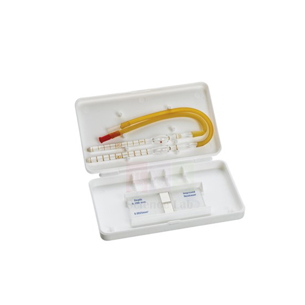
Haemocytometer
Haemocytometer (Product Code: SLE/M/88) is a glass counting chamber used to count cells in a diluted sample under a microscope in biology, microbiology and medical-laboratory teaching. This set has an improved Neubauer double-ruling counting chamber and comes with a pair of cover glasses and RBC and WBC diluting pipettes, so a class can dilute, load and count from one kit. It is supplied by Jain Scientific Equipments Pvt Ltd (ScienceLab), Ambala, India, for haemocytometer use in India’s labs and abroad.
Key Details
| Parameter | Detail |
|---|---|
| Chamber ruling | Improved Neubauer |
| Chamber type | Double ruling counting chamber |
| Cover glasses | A pair, included |
| Diluting pipettes | RBC and WBC diluting pipettes, included |
| Product code | SLE/M/88 |
| Supplied by | Jain Scientific Equipments Pvt Ltd (ScienceLab), Ambala, India |
What Is the Haemocytometer Used For?
In teaching laboratories the haemocytometer is the classic tool for learning manual cell counting. Students practise red and white blood cell counts, count yeast cells in fermentation experiments, or estimate the density of a cell or spore suspension in microbiology and cell-biology practicals. The two ruled areas of a double-ruling chamber allow two loadings to be counted and compared, a simple check on technique.
The general method is to dilute the sample with the matching pipette (the RBC pipette for red-cell work, the WBC pipette for white-cell work), place the cover glass over the ruled area, let the diluted sample run under it by capillary action, and count the cells in the defined squares under the microscope before calculating the concentration with the dilution used. Rinse the chamber and cover glasses with distilled water straight after use, dry them gently with lens tissue, and follow your laboratory’s biosafety procedure whenever the sample is blood. It is a core item for cell counting with a hemocytometer in India’s college labs.
What to Specify When Ordering
- Number of complete sets (chamber, cover glasses and both diluting pipettes).
- Whether spare cover glasses should be quoted for class use.
- Any microscopes or cell counters to add to the same enquiry.
Supplier
The Haemocytometer is supplied by Jain Scientific Equipments Private Limited, which operates under the ScienceLab name in Ambala, Haryana. We source this counting chamber set for colleges, schools, training institutes and laboratory dealers, and can include it with microscopes and cover glasses in a single quotation. Company background is available on our profile page.
Related Microscope Products
Items used in the same cell-counting practical:
- Cover Glasses — spares for class use and breakages.
- Student Medical Microscope — a microscope for counting under the ruled grid.
- Binocular Biological Microscope — comfortable two-eye viewing for longer counts.
- Manual Differential Cell Counter — for tallying cells as they are counted.
- Lens Tissue — for drying the chamber without scratching it.
See more in our Microscope category and the Clinical Laboratory range.
Frequently Asked Questions
What is included with the Haemocytometer (SLE/M/88)?
The Haemocytometer (SLE/M/88) includes an improved Neubauer double-ruling counting chamber, a pair of cover glasses, and RBC and WBC diluting pipettes. Together these let a student dilute a sample, load the chamber and count cells under a microscope.
What does double ruling mean on a haemocytometer?
A double-ruling Haemocytometer has two ruled counting areas on the same chamber. Students can load two samples, or the same sample twice, and compare the counts, which is a useful check on dilution and loading technique.
Which counting ruling does this Haemocytometer use?
This Haemocytometer uses the improved Neubauer ruling. Laboratory manuals and counting procedures written for the improved Neubauer pattern can therefore be followed directly, which keeps class instructions simple and consistent.
How should the Haemocytometer be cleaned after use?
Rinse the Haemocytometer chamber and cover glasses with distilled water immediately after use, then dry them gently with lens tissue. Avoid abrasive cloths and strong solvents that could damage the ruled surface, and handle the chamber only by its edges.
Is a calibration certificate available for the Haemocytometer?
Calibration or traceability documents for the Haemocytometer are not listed on this page. If your laboratory needs one, please discuss it with us through the contact page before ordering, and we will tell you what can be provided with the batch quoted.
Who supplies the Haemocytometer (SLE/M/88)?
Jain Scientific Equipments Pvt Ltd (ScienceLab), Ambala, India, supplies the Haemocytometer to colleges, schools, training institutes and laboratory dealers in India and international markets, and can quote it together with microscopes and cover glasses.
Request a Quote
Request the Haemocytometer (SLE/M/88) by sending the number of sets and your delivery details through our contact page, or add it to the enquiry cart with other lab items.
Last updated: 24 September 2026