- Physics Lab Equipment
- Chemistry Lab Equipment
- Biology Lab Equipment
- Anatomical Lab Equipment
- Learning
- Educational Lab Equipment
- Magnets
- Agriculture
- Astronomy
- Earth Science
- Geography Lab Equipment
- Safety Lab Coat
- Laboratory
- Meteorological
- Electricity & Magnetism
- Heat
- School Lab Equipment
- Lab Glassware
- Science Lab Equipment
- Tvet Lab Equipment
- Robotics Kits Supplier and Manufacturer for Schools India
- Math Lab Equipment Manufacturers and Exporters in Ambala
- STEM Kits Manufacturers and Supplier India
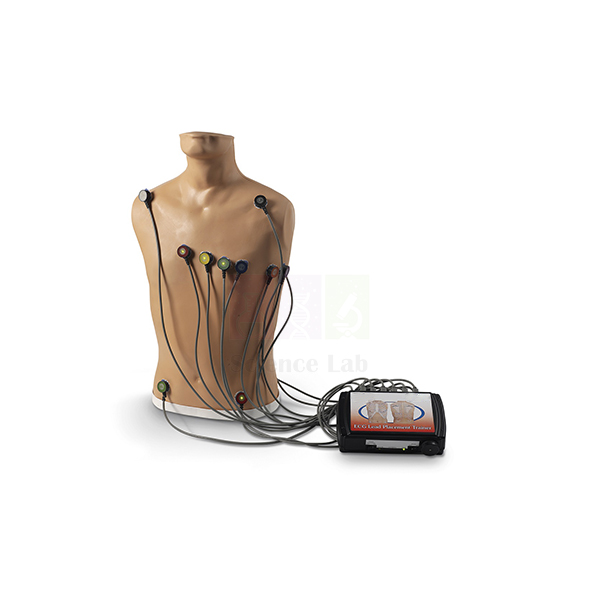
15-Lead ECG Placement Trainer
Teaches up to 15-lead ECG electrode placement anatomically and the accuracy of the electrode placement.
Students learn the placement of the electrodes on the adult trainer using anatomical landmarks, such as intercostal spaces, midclavicular line, anterior axillary line, midaxillary line, and scapula.
The trainer does not provide ECG output signals, but simulations of rhythms and hands-free defibrillation can be performed by adding any ECG arrhythmia simulator directly to an ECG monitor.
Connection sites for 4 limb leads and V1 through V9, with the ability to attach right- or left-sided electrodes.
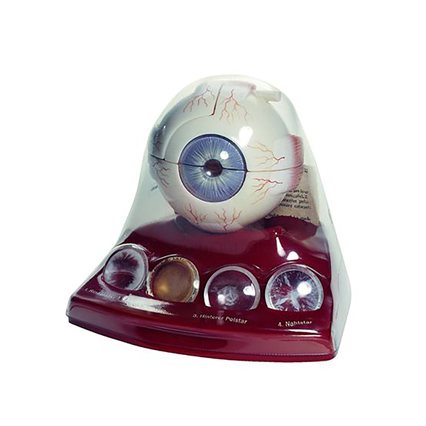
Cataract Eye Model with 4 Forms of Cataract
About 3x life size.
Size: 13 x 16 x 15 cm.
Model includes 4 forms of cataract: cortical, nuclear, posterior, and coronary.
Eye is mounted on base and forms of cataract rest in labeled compartments at bottom of base.
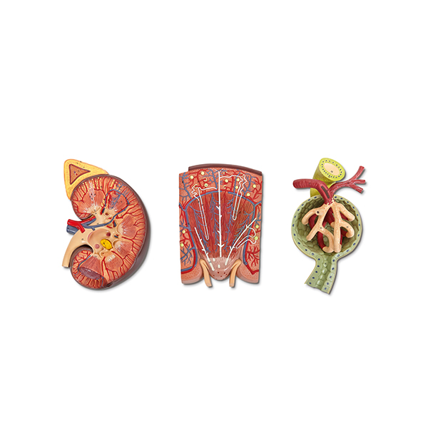
Human Kidney, Nephron and Renal Corpuscle Model
Greatly enlarged.
Size (overall), 69 × 29 × 10 cm.
Plaque-mounted series of 3 models, including a 2 1/2× life-size dissection of the kidney and adrenal gland.
Schematic enlargement showing anatomical details of an individual nephron; and a renal corpuscle with Bowman's capsule, glomerulus, and juxtaglomerular apparatus.
Contact Science Lab for your Educational School Science Lab Equipments. We are best educational lab equipments manufactruers, educational lab instruments manufacturer, educational laboratory glassware exporter, educational laboratory microscopes, educational laboratory suppliers, educational scienitific lab equipments.
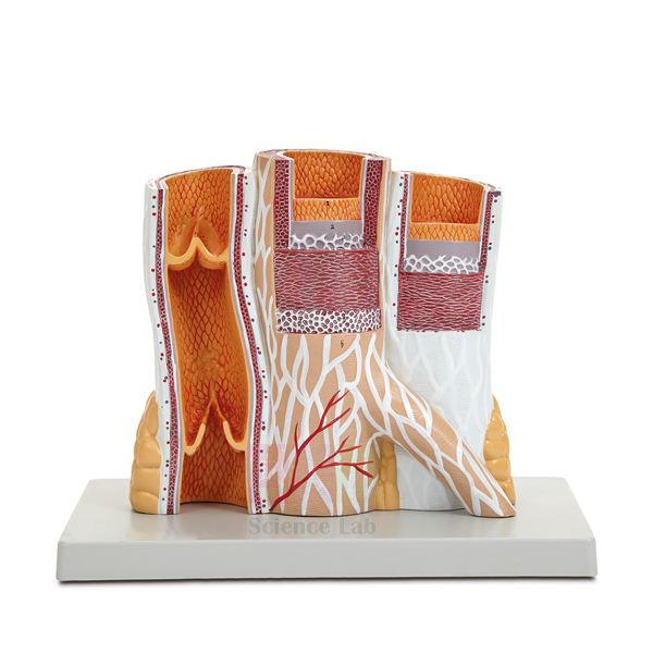
Microstructure of Artery and Vein Model
Includes numbered key.
Mounted on a durable polymer base.
Size (without base), 25 x 10 x 23 cm.
Included is a cut section of vein with venous valves.
Enlarged block model shows anatomical details of artery and vein.
Serial sectioning exposes sequential muscular and membranous layers and illustrates the differences between arterial and venous wall thickness.
Contact Science Lab for your Educational School Science Lab Equipments. We are best engineering educational equipments, high school science lab equipment manufacturers, lab equipment exporters, lab equipments manufactruers, lab instruments manufactruers, mathematics lab equipments manufacturers.
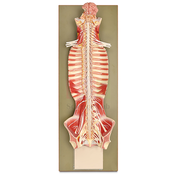
Human Nervous System Model
Life size.
On base. Size, 32 × 19 × 90 cm.
The sympathetic trunk is shown on left side.
Brain, brain stem, spinal cord, and spinal nerves are shown in position in vertebral column.
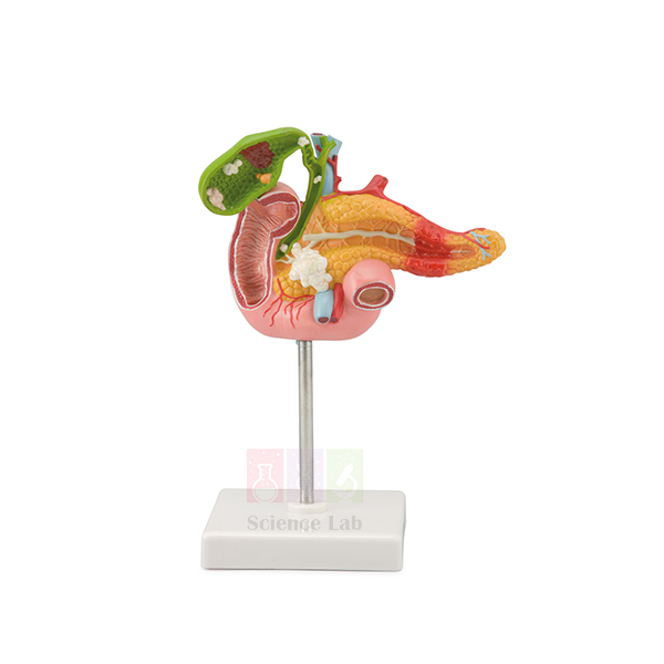
Pathology of the Human Pancreas, Duodenum, and Gallbladder Model
Life size.
On a base.
Size (with base), 11 x 11 x 24 cm.
This model depicts the most important diseases of the gallbladder, pancreas, and duodenum.
The gallbladder, partially opened, shows stones located in various locations, cholecystitis, polyposis, and carcinoma.
Diseases of the pancreas shown include pancreatitis in the pancreas tail and cancer in the pancreas head.
Also shown is a section of the duodenum with a duodenal ulcer.
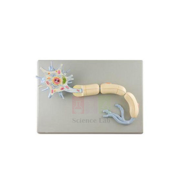
Neuron Model
About 2,500x life size.
Includes numbered key.
Size: 53 x 38 x 17 cm.
Mounted on a durable polymer base.
Detailed anatomical structures of a typical myelinated neuron are shown, including the cell body with nucleus and nucleolus; dendrites and axon; Golgi complex; mitochondria; endoplasmic reticulum; and myelin sheath with nodes of Ranvier and Schwann cells. Remove a portion of the myelin sheath to view the interior of the axon.
Contact Science Lab for your Educational School Science Lab Equipments. We are best scientific lab equipments manufacturers, scientific laboratory equipments manufacturer, technical educational equipment manufacturer, technical lab equipments manufacturers, tvet lab equipment manufacturers, vocational training lab equipments exporter.
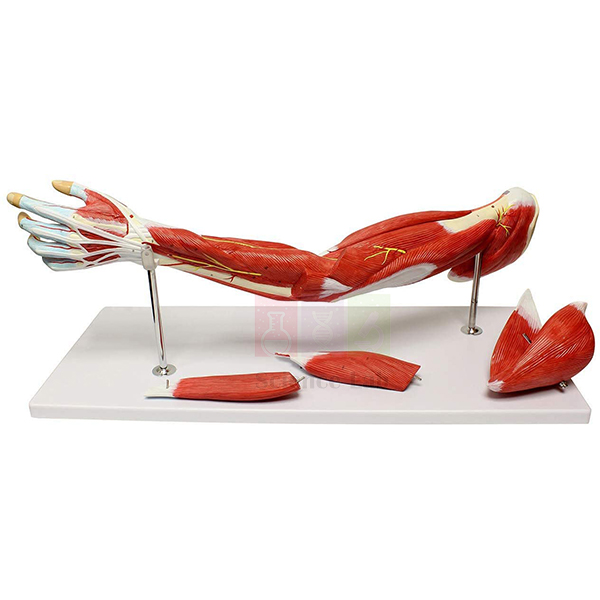
Human Muscular Arm Model
Life size.
Dissectible into 7 parts.
Size, 74 × 18 × 11 cm.
Muscles of the shoulder, arm, and hand are shown in great detail along with significant nerves and blood vessels.
Contact Science Lab for your Educational School Science Lab Equipments. We are best anatomy model manufacturer, biology lab equipment manufacturer, biology lab instruments exporter, biology lab instruments manufacturer, biology lab instruments supplier, burette pinchclip medium wall india.