- Physics Lab Equipment
- Chemistry Lab Equipment
- Biology Lab Equipment
- Anatomical Lab Equipment
- Learning
- Educational Lab Equipment
- Magnets
- Agriculture
- Astronomy
- Earth Science
- Geography Lab Equipment
- Safety Lab Coat
- Laboratory
- Meteorological
- Electricity & Magnetism
- Heat
- School Lab Equipment
- Lab Glassware
- Science Lab Equipment
- Tvet Lab Equipment
- Robotics Kits Supplier and Manufacturer for Schools India
- Math Lab Equipment Manufacturers and Exporters in Ambala
- STEM Kits Manufacturers and Supplier India
- Engineering Lab Equipment
- Hospital Lab Equipment
- Water, Sanitation & Hygiene Supplies
- Cold Chain, Solar & Communication Equipment
- Nutrition
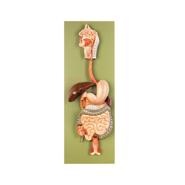
Human Digestive Tract Model
Human Digestive Tract Model (Product Code: SLE/A/99) presents the human digestive tract at life size on a single base, with a stomach that can be opened and a design that dissects into 3 pieces. It gives biology classes, nursing students and nutrition trainers a full-scale reference for how food travels through the body. The model is manufactured by Jain Scientific Equipments Pvt Ltd (ScienceLab), Ambala, a biology lab equipment supplier in India serving schools, colleges and training institutes.
Specifications
| Parameter | Detail |
|---|---|
| Type | Model of the human digestive tract |
| Scale | Life size |
| Dissection | Dissectible into 3 pieces |
| Stomach | Can be opened to view the interior |
| Size | 32 × 12 × 91 cm |
| Mounting | On base |
Typical Use
In a digestion lesson the teacher can start at the top of the model and move downward, naming each stage of the journey and explaining what happens to food there. Opening the stomach turns an abstract idea into something visible: students can discuss how the stomach stores and churns food and why its lining matters, then close it again and continue along the tract. Because the model is life size, learners come away with a realistic sense of how long the digestive route is.
The model suits nutrition and digestion chapters in senior school, introductory anatomy for nursing and allied-health courses, and community health talks. It is a staple of high school biology lab equipment for this reason. With a longest dimension of 91 cm, it needs a stable table or a dedicated display space; move it by the base rather than by the organs, and keep the 3 pieces together between classes.
Manufacturer
ScienceLab is the brand name of Jain Scientific Equipments Private Limited, which manufactures this digestive tract model and runs its business from Ambala in the state of Haryana. The company equips laboratories in India and international markets; the company profile gives an overview of its work.
Related Human Models
Pieces that extend a digestive system lesson:
- Human Stomach Model — a closer look at the organ that opens on this model
- Human Liver and Gallbladder Model — the accessory organs that supply bile to the tract
- Human Spleen, Duodenum, and Pancreas Model — detail of the region just beyond the stomach
- Human Digestive System Model — an alternative model of the digestive system for comparison
- Microslide The Digestive System — prepared slides for the microscopic view of digestive tissues
For more anatomy teaching models, browse the Human Models category.
Frequently Asked Questions
How many pieces does it separate into?
Three. The model is dissectible into 3 pieces, and the stomach can also be opened.
How much space does it need?
It measures 32 × 12 × 91 cm on its base, so plan for a sturdy table or cabinet shelf that can take the 91 cm dimension.
Which organs are represented?
It represents the human digestive tract at life size, including an opening stomach. For a structure-by-structure list, please contact us before ordering.
Can it be combined with microscope work?
Yes. Many teachers follow the model with prepared digestive-system slides so students see the same tract at tissue level; see the slide set in the related list.
How do we keep it in good condition?
Dust it regularly, wipe marks with a slightly damp soft cloth, and store it away from direct sunlight and heat. Reassemble the pieces fully before moving it.
What warranty is offered?
Warranty terms are confirmed at the quotation stage; contact us for details.
Can you supply it in quantity for several labs?
Yes. We supply schools, colleges and distributors in India and international markets; include the quantity and delivery location in your enquiry cart request.
Request a Quote
Ask for a price on the Human Digestive Tract Model via our contact page, or add it to the enquiry cart with related digestive system models.
Last updated: 24 September 2026
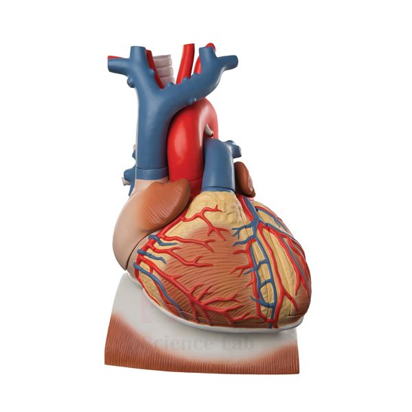
Human Heart on Diaphragm Model
Human Heart on Diaphragm Model (Product Code: SLE/A/100) shows a life-size human heart resting on the diaphragm, the way it sits in the chest, and dissects into 8 parts that include the diaphragm with a section of pericardium, the thymus gland and the ventricles. It helps students connect cardiac anatomy with the heart's position and surroundings, making it a strong heart model for anatomy teaching in India's schools, nursing colleges and medical institutes. It is manufactured by Jain Scientific Equipments Pvt Ltd (ScienceLab), Ambala.
Specifications
| Parameter | Detail |
|---|---|
| Type | Heart model mounted on the diaphragm |
| Scale | Life size |
| Dissection | Dissects into 8 parts |
| Parts include | Diaphragm with section of pericardium, thymus gland, apex of the heart, lower part of the ventricles, and ventricles |
| Size | 29 x 18 x 19 cm |
| Mounting | On a green base |
Typical Use
Most heart models show the organ on its own; this one keeps it on the diaphragm so a lesson can begin with position. Students see that the heart rests on the muscular floor of the chest, wrapped by the pericardium, with the thymus gland in front of it. The teacher can then lift away the thymus and open the lower heart by removing the apex and ventricle sections, moving from surface anatomy to the internal chambers in one sequence.
This makes it useful when teaching breathing and circulation together, since the diaphragm's role in respiration can be discussed right beside the heart it supports. It is a sensible centrepiece for a school biology lab setup in India and for first-year anatomy practicals. After each class, check that all 8 parts are back in place so none are lost between sessions.
Manufacturer
Made by Jain Scientific Equipments Private Limited, the Ambala, Haryana company behind the ScienceLab brand, this model belongs to a wider line of anatomical teaching models supplied to institutions in India and international markets. See the company profile for more about the manufacturer.
Related Human Models
Other models that support cardiovascular and thoracic lessons:
- Human Heart Model — a stand-alone heart for studying external features
- Heart 3 times Natural Size Model — an enlarged heart for viewing detail from a distance
- Human Lungs with Heart and Larynx Model — shows the heart together with the lungs and airway
- Human Thoracic Organs Model — the wider chest cavity in which heart and diaphragm sit
- Microslide The Circulatory System — prepared slides that take circulation lessons to tissue level
Browse the complete Human Models range for further options.
Frequently Asked Questions
Which parts come off the model?
It dissects into 8 parts, including the diaphragm with a section of pericardium, the thymus gland, the apex of the heart, the lower part of the ventricles and the ventricles. For the complete part list, contact us.
Why is the heart shown on the diaphragm?
It keeps the heart in its natural relationship with the diaphragm and pericardium, so students learn position and surroundings as well as the organ itself.
What are its size and scale?
The model is life size and measures 29 x 18 x 19 cm on a green base.
Is there a numbered key?
A key is not mentioned in our product details for this model; please ask us to confirm what is supplied.
Which classes is it best for?
It suits senior school biology, nursing and paramedical anatomy, and introductory medical anatomy where heart position and structure are taught together.
How should it be looked after?
Handle the removable parts over a table, wipe with a soft, lightly damp cloth, and store the assembled model covered to keep dust off.
How do we order several units?
Send your quantity and delivery location through the contact page or enquiry cart; we supply institutions and distributors in India and international markets.
Request a Quote
For a quotation on the Human Heart on Diaphragm Model, contact our team or add the model to your enquiry cart.
Last updated: 24 September 2026
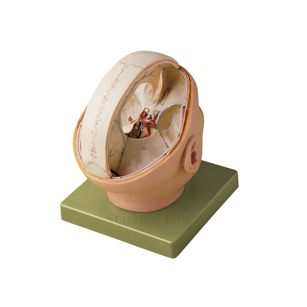
Human Dura Mater Model
Human Dura Mater Model (Product Code: SLE/A/101) is a life-size neuroanatomy model that shows the dura mater with its sinuses, the 12 pairs of cranial nerves and the arteries at the base of the brain, and separates into 2 parts for closer study. Anatomy lecturers, nursing tutors and senior biology teachers use it to explain how the brain is protected, supplied with blood and connected to the head and neck. It is manufactured by Jain Scientific Equipments Pvt Ltd (ScienceLab), an anatomical models manufacturer in Ambala.
Specifications
| Parameter | Detail |
|---|---|
| Type | Neuroanatomy model of the dura mater |
| Scale | Life size |
| Dissection | Dissectable into 2 parts |
| Size | 18 × 21 × 23 cm |
| Mounting | On base |
| Structures shown | Dura mater with its sinuses in proportion, the 12 pairs of cranial nerves, and the arteries at the base of the brain |
Typical Use
The model is built for lessons on the meninges and the base of the brain. A lecturer can first show the dura mater as the tough outer covering and point out its sinuses, which collect blood leaving the brain, then turn the model over to trace the 12 pairs of cranial nerves and the arteries that run along the underside. Students revising the cranial nerves often find it easier to learn their order and position on a three-dimensional model than from a textbook drawing.
It is used in human anatomy and physiology practicals at medical, dental, nursing and physiotherapy colleges, and in advanced school biology when the nervous system is covered in depth. Institutions expanding their educational laboratory equipment in India often add it alongside a brain model and a skull, so the brain, its coverings and the bony case can be taught as one topic. Set it on a table at student eye level when discussing the base-of-brain structures.
Manufacturer
Jain Scientific Equipments Private Limited manufactures the Human Dura Mater Model and sells it under its trading name, ScienceLab. The company is headquartered in Ambala, Haryana, and supplies anatomy and laboratory products to customers across India and international markets. Visit the company profile to learn more.
Related Laboratory Equipment
Models that complete a lesson on the brain, its coverings and its nerves:
- Human Brain Model with Arteries — follows the arterial supply of the brain in more detail
- Human Brain Stem Model — shows the region from which most cranial nerves emerge
- Human Brain Model, 15 Parts — a dissectible brain for the structures beneath the dura
- Human Skull Model — the bony case that the dura mater lines
- Human Nervous System Model — carries the nerve pathways beyond the skull
Find more options in our Human Models category.
Frequently Asked Questions
What anatomy does the model display?
The dura mater with its sinuses, the 12 pairs of cranial nerves, and the arteries at the base of the brain, all at life size.
Into how many parts does it separate?
Two. It is dissectable into 2 parts so both the covering and the underlying structures can be examined.
What are the dimensions?
It measures 18 × 21 × 23 cm and is supplied on a base.
Are the cranial nerves labelled or numbered?
All 12 pairs are shown, but a numbered key is not listed in our product details. Please contact us to confirm labelling before you order.
Is it suitable for school use as well as colleges?
Yes, although it is most often chosen for college-level anatomy; senior school classes use it when studying the nervous system in depth.
How are warranty and after-sales questions handled?
Warranty terms are set out in the quotation. Reach out with any after-sales query.
What should an institutional enquiry include?
Quantity, delivery location and any companion models you need. Submit it via the contact form or the enquiry cart.
Request a Quote
Prices and availability for the Human Dura Mater Model are available on request: contact us or use the enquiry cart.
Last updated: 24 September 2026
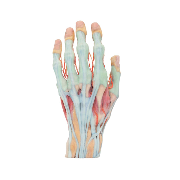
3-D Printed Human Superficial Hand Dissection Model
3-D Printed Human Superficial Hand Dissection Model (Product Code: SLE/A/102) reproduces a superficial dissection of the human hand at about life size, showing the vasculature, musculature and tendons in intricate detail. Because it is 3-D printed, students can study a specimen that is as realistic as possible without the need for human cadavers. It is used in anatomy, physiotherapy and occupational therapy teaching, and is one of the human anatomical models manufactured in India by Jain Scientific Equipments Pvt Ltd (ScienceLab), Ambala.
Specifications
| Parameter | Detail |
|---|---|
| Type | 3-D printed superficial dissection model of the human hand |
| Scale | About life size |
| Size | About 6-7/10" W x 5-9/10" H x 11-2/5" L |
| Structures shown | Vasculature, musculature and tendons of the hand, in intricate detail |
| Purpose | Realistic specimen study without the need for human cadavers |
Typical Use
The hand packs a great deal of anatomy into a small space, and this model lets learners examine the layer just beneath the skin: the blood vessels that supply the fingers and palm, the small muscles that give the hand its precision, and the tendons that transmit movement from the forearm. Instructors use it to explain grip, pinch and finger movement, and to show why injuries to a single tendon or vessel can affect function so noticeably.
For courses where cadaver access is limited, or where the biology dissection set is used only on animal specimens, the model gives a realistic human reference that can be handled in every session without preservation or special storage. Physiotherapy, occupational therapy, nursing and medical students use it for hand-anatomy practicals, and art and design departments use it to study the hand's form. Pairing it with a skeletal hand helps students relate each tendon to the bones it moves.
Manufacturer
The model is produced by Jain Scientific Equipments Private Limited, which trades as ScienceLab from its base in Ambala, Haryana, and serves teaching institutions in India and international markets. Buyers who need company registration details for procurement can request them with their quotation. The company profile has further background.
Related Human Models
Models that help build a complete picture of the hand and upper limb:
- Human Skeletal Hand Model — the bones beneath the muscles and tendons shown here
- Human Hand Joint Model with Ligaments — adds the joints and ligaments of the hand
- Human Arm and Hand model — follows the tendons back to the forearm muscles
- 3-D Printed Internal Heart Structures Model — another 3-D printed model for cadaver-free study
- 3D Printed Human Flexed Knee Joint — a 3-D printed joint for lower-limb lessons
See all Human Models for more anatomy teaching options.
Frequently Asked Questions
What does "superficial dissection" mean on this model?
It shows the structures that lie just beneath the skin of the hand: the vasculature, musculature and tendons.
How large is it?
About life size, measuring about 6-7/10" W x 5-9/10" H x 11-2/5" L.
Why choose a 3-D printed model over a cadaveric specimen?
It lets students study a realistic specimen without the need for human cadavers, so it can be used in ordinary classrooms and handled repeatedly.
Are the bones of the hand shown?
Our product details describe the vasculature, musculature and tendons. For bone anatomy, add the Human Skeletal Hand Model, or ask us about the model's deeper layers.
Does it come on a stand or base?
A stand is not listed for this model. Please contact us to confirm how it is supplied.
How should it be cleaned?
Dust it with a soft dry brush or cloth, and use only a lightly damp cloth for marks; avoid solvents and hot water.
What information speeds up a quotation?
The number of units, your delivery location and any registration documents your purchasing office requires. Send these via the contact page or the enquiry cart.
Request a Quote
To order the 3-D Printed Human Superficial Hand Dissection Model, contact us for pricing or add it to your enquiry cart.
Last updated: 24 September 2026
Human Brain with Cortical Functional Centers Model
Human Brain with Cortical Functional Centers Model (Product Code: SLE/A/103) is a life-size, 15-part dissectible brain used to teach the localisation of function in the cerebral cortex in psychology, neuroscience, medical and nursing courses. Its left hemisphere is colour coded with the official numbers of Brodmann’s cytoarchitectural map, and the brain rests in a lifelike position in a skull base mounted on a stand. It is made by Jain Scientific Equipments Pvt Ltd (ScienceLab), an OEM human anatomical models manufacturer, at its own factory in Ambala, India.
Specifications
| Parameter | Detail |
|---|---|
| Type | Dissectible functional brain model in skull base |
| Scale | Life size |
| Parts | Dissectible into 15 parts |
| Size | 23 x 15 x 18 cm |
| Mounting | Rests in a lifelike position in the base of a skull, mounted on a stand |
| Left hemisphere | Colour coded and marked with the official numbers of Brodmann’s cytoarchitectural map of the cerebral cortex |
| Highlighted areas | Primary and secondary motor and sensory cortical fields; areas associated with speech, audition and vision; structures of the limbic system |
| Product code | SLE/A/103 |
| Manufacturer | Jain Scientific Equipments Pvt Ltd (ScienceLab) — OEM |
| Country of origin | India (made in Ambala, Haryana) |
What Is the Cortical Functional Centers Brain Model Used For?
A lecture on localisation of function becomes much clearer when students can hold the brain and see the numbered Brodmann areas on the left hemisphere. A tutor might start with the primary motor and sensory fields, move to the speech areas, then show the auditory and visual regions, asking learners to predict which abilities are affected by damage to each area. Taking the brain apart into its 15 parts then reveals the deeper limbic structures involved in memory and emotion.
Seating the brain back in the skull base shows how it sits within the cranial cavity, which helps when discussing head injuries and the position of cortical areas under the skull. Psychology departments and medical colleges buying biology teaching aids in India value this combination of structure and function in one model.
OEM Manufacturer
Behind this functional brain is its original equipment manufacturer (OEM): Jain Scientific Equipments Private Limited, which trades as ScienceLab and makes the complete model at its own licensed factory at Saha, Ambala, Haryana, India.
Psychology departments, medical colleges and distributors order it factory-direct, and questions about a replacement part or a repeat order are answered by the manufacturer — ask us. Learn more on the company profile page.
Related Brain Models
Consider these alongside the functional brain for a complete neuroscience teaching set:
- Human Brain Model, 15 Parts — an alternative dissectible brain for general anatomy classes
- Human Regional Brain Model — a simpler two-part brain colour coded by functional area
- Human Brain Model with Arteries — links cortical areas to their blood supply, useful when teaching stroke
- Human Brain Stem Model — covers the region that connects the cortex with the spinal cord
- Human Nervous System Model — places the brain within the whole nervous system
The full Brain Models collection lists every option.
Frequently Asked Questions
Which hemisphere of the functional centers brain shows the Brodmann numbers?
On the Human Brain with Cortical Functional Centers Model, the left cerebral hemisphere is colour coded and marked with the official numbers of Brodmann’s cytoarchitectural map of the human cerebral cortex.
How many parts does the cortical functional centers brain separate into?
The Human Brain with Cortical Functional Centers Model is dissectible into 15 parts, which allows the deeper limbic structures and other internal parts to be examined after the cortical areas have been studied.
Is a full skull included with this brain model?
The brain of the Human Brain with Cortical Functional Centers Model rests in the base of a skull mounted on a stand. A full skull cap is not listed in our product information, so contact us if you need one.
What are the dimensions of the Human Brain with Cortical Functional Centers Model?
The Human Brain with Cortical Functional Centers Model measures 23 x 15 x 18 cm, and the brain itself is life size, so it can be compared directly with specimens, atlases and scan images.
Is the functional centers brain useful for psychology rather than medicine?
Yes. The highlighted speech, hearing, vision, motor and sensory areas and the limbic structures on the Human Brain with Cortical Functional Centers Model match topics taught in biological psychology and neuroscience courses as well as medicine.
Who manufactures the Human Brain with Cortical Functional Centers Model?
The Human Brain with Cortical Functional Centers Model is manufactured by Jain Scientific Equipments Pvt Ltd (ScienceLab), the OEM, at its own licensed factory at Saha, Ambala, Haryana, India, and is supplied factory-direct to institutions and dealers.
What should a tender or purchase request include?
A tender or purchase request for the Human Brain with Cortical Functional Centers Model should quote product code SLE/A/103, the quantity, the delivery address and any documents your procurement office needs; send it through our contact page.
Request a Quote
For pricing and availability of this functional brain model, contact us via the enquiry form or add it to your enquiry cart with other anatomy models. The quotation is issued by the manufacturer.
Last updated: 24 September 2026
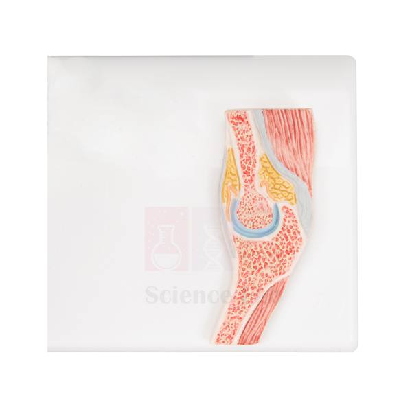
Human Elbow Cross-Section Model
Human Elbow Cross-Section Model (Product Code: SLE/A/104) is an intricately crafted, life-size section through the human elbow in which the muscles, ligaments and bones are clearly shown. Important structures are numbered against a key printed on the green base, and a removable transparent plastic cover protects the section. Anatomy, physiotherapy and sports-science teachers use it to explain how the elbow is built in layers. It is manufactured by Jain Scientific Equipments Pvt Ltd (ScienceLab), a laboratory equipment supplier in India based in Ambala.
Specifications
| Parameter | Detail |
|---|---|
| Type | Cross-section model of the human elbow |
| Scale | Life size |
| Size | 32 x 26 x 4 cm |
| Structures shown | Muscles, ligaments and bones |
| Identification | Important structures numbered; key printed on the green base |
| Protection | Removable, transparent plastic cover |
Typical Use
A cross-section answers a question that whole-limb models leave open: what lies next to what at one level of the arm. With this model, students can see how muscle groups wrap around the bones of the elbow and where the ligaments hold the joint together, then use the numbered key to name each structure. It works well for self-study, because learners can write down the numbers on a worksheet and check their answers against the printed key.
Used next to a human skeleton model, it helps students relate the flat section back to the whole arm and understand how injuries around the elbow affect nearby tissues. The slim 4 cm depth and protective cover make it practical for display cabinets, anatomy museums and corridor showcases as well as bench teaching; keep the cover in place whenever the model is not being examined.
Manufacturer
Behind the ScienceLab name is Jain Scientific Equipments Private Limited, an Ambala, Haryana company that manufactures this elbow section among its many anatomical and laboratory products for buyers in India and international markets. Learn more on the company profile page.
Related Human Models
Models that pair with this section for upper-limb teaching:
- Human Elbow Joint Model — the complete joint for comparing with the sectional view
- Functional Elbow Joint — demonstrates how the joint moves, which a section cannot show
- Human Muscular Arm Model — follows the muscles seen here along the full arm
- Shoulder Girdle with Musculature Model — continues the upper limb up to the shoulder
- Human Skeleton, Rod-Supported — a full skeleton for placing the elbow in context
More anatomy models are listed in the Human Models category.
Frequently Asked Questions
Does it come with a protective cover?
Yes. A removable, transparent plastic cover is included to protect the section.
How are the structures identified?
Important structures are numbered on the model, and the key is printed on the green base.
What are its dimensions?
The model is life size and measures 32 x 26 x 4 cm.
Can it demonstrate elbow movement?
No. It is a fixed cross-section for studying structure; for flexion and extension, see the Functional Elbow Joint in the related list.
How should the model and cover be cleaned?
Wipe the cover with a soft, lint-free cloth, and dust the model only when the cover is off. Avoid solvent-based cleaners on the transparent plastic.
What is the warranty?
Warranty terms are confirmed in the quotation; contact us for current terms.
Can it be supplied in multiples or exported?
Yes. Multiple units can be supplied to institutions and distributors in India and international markets; send your requirement through the enquiry cart or contact page.
Request a Quote
For a price on the Human Elbow Cross-Section Model, send us an enquiry or add it to the enquiry cart.
Last updated: 24 September 2026
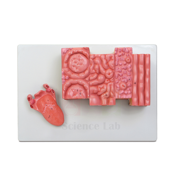
Microstructure of the Human Tongue Model
Microstructure of the Human Tongue Model (Product Code: SLE/A/105) enlarges a block of tongue tissue about 20x so that taste buds, lingual and mucous glands and the fine structure of the musculature can be seen without a microscope. A separate life-sized model of the intact tongue, with the palatine and lingual tonsils and the epiglottis, is included for orientation. Histology lecturers and biology teachers use the pair to link tissue structure with the whole organ. It is manufactured by Jain Scientific Equipments Pvt Ltd (ScienceLab), a biology lab instruments supplier in Ambala.
Specifications
| Parameter | Detail |
|---|---|
| Type | Enlarged microstructure model of the human tongue |
| Scale | About 20x life size |
| Size | 33 × 23 × 11 cm |
| Mounting | Durable polymer base |
| Histological features | Taste buds, lingual and mucous glands, fine structure of the musculature |
| Key | Numbered key included |
| Also included | Separate life-sized intact tongue model featuring the palatine and lingual tonsils and the epiglottis |
Typical Use
This model is designed for teaching at two scales. Start with the life-sized tongue to orient the class: where the tonsils sit, how the epiglottis relates to the back of the tongue, and why this region matters for swallowing. Then move to the enlarged block, where students can find the taste buds, follow the glands that keep the mouth moist, and see how muscle fibres run in different directions to give the tongue its flexibility.
It is especially helpful just before a practical with prepared microscope slides, because students who have already seen the structures in three dimensions recognise them far more quickly under the lens. Dental, medical, nursing and food-science courses use it in oral histology and taste physiology, and senior school classes use it in lessons on the senses. The numbered key makes it easy to set identification exercises.
Manufacturer
Jain Scientific Equipments Private Limited, trading as ScienceLab, manufactures this tongue microstructure model. The company is located in Ambala, Haryana, and provides histology, anatomy and general laboratory products to education buyers in India and international markets. Its background is summarised on the company profile.
Related Laboratory Equipment
Items that support an oral histology or taste lesson:
- Human Tongue Model — an additional whole-tongue model for group work
- Prepared Microscope Slide Set – Digestion (Liver, Pancreas And Tongue) — real tongue tissue to examine after the model
- Microslide Basic Human Histology — broader histology slides for comparing tissue types
- Compound Microscope — for viewing the slides that follow the model demonstration
- Larynx Human Model — continues from the epiglottis into the larynx
Explore more anatomy models in the Human Models category.
Frequently Asked Questions
What is included in the set?
The enlarged microstructure model on its polymer base, a numbered key, and a separate life-sized model of the intact tongue.
How much is the tissue enlarged?
About 20x life size; the enlarged model measures 33 × 23 × 11 cm.
Which microscopic features can students identify?
Taste buds, lingual and mucous glands, and the fine structure of the tongue musculature.
What does the life-sized tongue show?
The intact tongue with the palatine and lingual tonsils and the epiglottis, helping students place the enlarged tissue in context.
Can it replace microscope slides in a practical?
No, it complements them. The model prepares students to recognise structures, and slides then show real tissue.
How should it be cleaned?
Wipe the model and its polymer base with a soft, slightly damp cloth, then dry. Keep the life-sized tongue with the main model so it is not misplaced.
How can we get a quotation for several departments?
List the quantities and delivery addresses in the enquiry cart or on our contact page; warranty terms are included with the quotation.
Request a Quote
Pricing for the Microstructure of the Human Tongue Model is available on request — contact us or use the enquiry cart.
Last updated: 24 September 2026
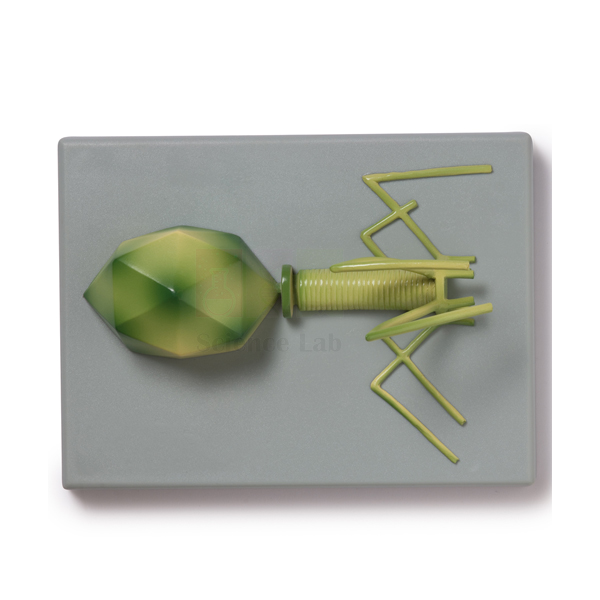
Bacteriophage T4 Model
Bacteriophage T4 Model (Product Code: SLE/A/106) is a one-piece teaching model of the T4 virus, enlarged millions of times, used to explain virus structure and how a phage attaches to the bacterium Escherichia coli in biology and microbiology classes. Mounted on a durable polymer base and supplied with a key, it helps school and college teachers make an invisible organism concrete. The model is made by Jain Scientific Equipments Pvt Ltd (ScienceLab), an OEM manufacturer, at its own factory in Ambala, India.
Specifications
| Parameter | Detail |
|---|---|
| Subject | Bacteriophage T4, a virus that attaches to the surface of the much larger bacterium Escherichia coli |
| Scale | Enlarged millions of times |
| Construction | One piece |
| Mounting | Durable polymer base |
| Supplied with | Key |
| Product code | SLE/A/106 |
| Manufacturer | Jain Scientific Equipments Pvt Ltd (ScienceLab) — OEM |
| Country of origin | India (made in Ambala, Haryana) |
What Is the Bacteriophage T4 Model Used For?
Viruses are hard to teach because students never see one. A solid model lets learners examine the phage's head, tail and tail fibres from every side while the teacher explains the stages of infection: attachment to the bacterial surface, injection of genetic material, copying of viral parts inside the host and release of new phages. Using the key, students can label a sketch of the model and write the infection sequence beside it, a useful revision exercise before exams.
The model also supports discussions of scale. Since T4 infects E. coli, a bacterium far smaller than an animal cell, comparing it with a cell model or a stained bacteria slide gives students a feel for how small viruses really are. As part of the microbiology lab equipment in a school or college collection, it suits units on microorganisms, disease, biotechnology and the nature of genetic material.
OEM Manufacturer
ScienceLab is the trading name of Jain Scientific Equipments Private Limited, the OEM that makes this bacteriophage model at its own licensed factory at Saha, Ambala, Haryana. The company is a registered MSME with manufacturing as its major activity (Udyam UDYAM-HR-01-0003714), and overseas consignments move under DGFT Importer-Exporter Code AAECJ8618A. Institutions, dealers and importers deal with the manufacturer directly for quotations and repeat orders; see the company profile.
Related Animal Models
Useful companions for a lesson on viruses and bacteria:
- Bacteria slide Set of 12 — real bacterial forms, including Escherichia coli, for students to observe after studying the phage's host.
- Monocular Microscope 1000x, LED light — the magnification needed to view some of the bacteria slides.
- Viruses & Bacteria Poster — a wall reference on common viruses and bacteria to display beside the model.
- DNA-RNA Model — builds the genetic material that a phage injects into its host.
Other biological models are listed in the Animal Model category.
Frequently Asked Questions
Does the Bacteriophage T4 Model come with a key?
Yes. The Bacteriophage T4 Model is supplied with a key, so students can identify the parts of the virus shown on the model and use the same names when they label their own diagrams or write out the stages of infection.
Does the Bacteriophage T4 Model come apart?
No. The Bacteriophage T4 Model is made as a single piece fixed to a durable polymer base, which keeps the whole structure secure when it is passed around a class or carried between teaching rooms.
Why use the Bacteriophage T4 Model instead of a microscope slide?
Viruses such as T4 are far too small for a school light microscope, so the Bacteriophage T4 Model, enlarged millions of times, is the practical way to show a virus's shape in class and compare it with the bacterium it infects.
Who manufactures the Bacteriophage T4 Model?
Jain Scientific Equipments Pvt Ltd (ScienceLab) manufactures the Bacteriophage T4 Model as the OEM at its own licensed factory at Saha, Ambala, Haryana, India. Schools, colleges and dealers can buy it factory-direct and receive quotations from the maker itself.
What are the overall dimensions of the Bacteriophage T4 Model?
The physical size of the Bacteriophage T4 Model is not listed in its product details. Ask us through the contact page and we will confirm the dimensions so you can plan cabinet or display space before ordering.
Which topics does the Bacteriophage T4 Model support?
The Bacteriophage T4 Model supports lessons on microorganisms, viral infection of bacteria, disease, biotechnology and DNA as genetic material, and it is a handy revision aid when students must describe the stages of phage infection.
Can the Bacteriophage T4 Model be supplied to colleges outside India?
Yes. The Bacteriophage T4 Model is supplied to institutions in India and international markets. Include the destination, quantity and any import documents your college needs in the enquiry so the manufacturer can quote accordingly.
Request a Quote
Ask for pricing on the Bacteriophage T4 Model through our contact page, or collect it with other items in the enquiry cart and send one combined request; the quotation comes straight from the manufacturer.
Last updated: 24 September 2026