- Physics Lab Equipment
- Chemistry Lab Equipment
- Biology Lab Equipment
- Anatomical Lab Equipment
- Learning
- Educational Lab Equipment
- Magnets
- Agriculture
- Astronomy
- Earth Science
- Geography Lab Equipment
- Safety Lab Coat
- Laboratory
- Meteorological
- Electricity & Magnetism
- Heat
- School Lab Equipment
- Lab Glassware
- Science Lab Equipment
- Tvet Lab Equipment
- Robotics Kits Supplier and Manufacturer for Schools India
- Math Lab Equipment Manufacturers and Exporters in Ambala
- STEM Kits Manufacturers and Supplier India
- Engineering Lab Equipment
- Hospital Lab Equipment
- Water, Sanitation & Hygiene Supplies
- Cold Chain, Solar & Communication Equipment
- Nutrition
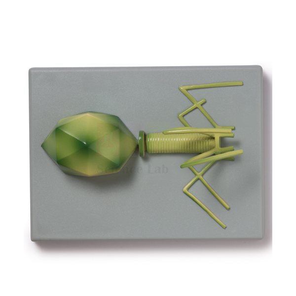
Bacteriophage T4 Model
The Bacteriophage T4 Model is a one-piece teaching model of the T4 virus, enlarged millions of times and fixed to a durable polymer base. T4 infects the bacterium Escherichia coli, and the model lets a class study the head, tail and tail fibres from every side. A key comes with it for naming the parts.
Bacteriophage T4 Model specifications
| Parameter | Detail |
|---|---|
| Subject | Bacteriophage T4, a virus that attaches to the much larger bacterium Escherichia coli |
| Scale | Enlarged millions of times |
| Construction | One piece |
| Mounting | Durable polymer base |
| Supplied with | Key |
| Product code | SLE/A/106 |
| Minimum order | 10 units |
| Dispatch | Usually within 7 to 10 working days |
| Manufacturer | ScienceLab (Jain Scientific Equipments Pvt Ltd), Ambala, India |
Teaching viruses with the T4 phage model
Labelling the infection sequence. Students sketch the model, name its parts from the key and write the stages of the lytic cycle beside the drawing: attachment, injection, copying of viral parts, assembly and release. The Viruses & Bacteria Poster on the wall gives a second reference while they work.
Host and virus. Students view rod-shaped bacteria from the bacteria slide set under a high-power LED microscope, then return to the model. The comparison makes the point that a phage is far smaller than its host and cannot be seen with a light microscope.
Virus versus cell. Set the model beside a prokaryotic cell model and list what the bacterium has that the phage lacks: a cell membrane, cytoplasm, ribosomes and its own metabolism. This answers the frequent mix-up between viruses and bacteria.
What gets injected. Build a strand on the DNA-RNA model to show the genetic material the phage delivers. Then discuss the classic blender experiment, run with a close relative of T4, which showed that DNA and not protein enters the host cell.
Frequently Asked Questions About the Bacteriophage T4 Model
What are the main parts of a T4 phage?
T4 has a many-sided protein head, the capsid, that holds its DNA, a tail with a contractile sheath, and long tail fibres that grip the host. Unlike influenza or HIV, it has no outer envelope. The key supplied with the model names each part shown.
Why is the head of the phage shaped like an icosahedron?
A shell built from many identical protein units closes most efficiently into a regular many-faced shape, so the virus needs few genes to make it. In T4 the head is an icosahedron stretched along one axis and packed tightly with DNA.
How does T4 get its DNA into the bacterium?
The tail fibres bind to receptors on the surface of E. coli and the base of the tail settles onto the cell wall. The sheath then contracts and drives the inner tube through the wall. The DNA passes into the cell while the empty protein coat stays outside.
Is a virus a living thing?
It is a borderline case. A phage carries genes and evolves, but it cannot grow, respire or copy itself outside a host. Most textbooks treat viruses as obligate intracellular parasites: inactive particles until they enter a suitable cell.
What is the difference between the lytic and lysogenic cycles?
In the lytic cycle the phage takes over the cell at once, makes new phages and bursts the cell to release them. In the lysogenic cycle the viral DNA joins the host chromosome as a prophage and is copied with it until a trigger switches it to the lytic route. T4 is a lytic phage.
Can a phage like T4 infect human cells?
No. A phage can multiply only inside bacteria whose surface receptors it recognises, so T4 attacks E. coli and cannot reproduce in human cells. For the same reason, phages are being studied as a way to kill bacteria that resist antibiotics.
Virus, bacteria and DNA teaching aids
- Bacteria slide Set of 12
- Viruses & Bacteria Poster
- DNA-RNA Model
- Prokaryotic Cell Model
- Microslide Helpful Bacteria
- Microslide Harmful Bacteria
To request a quotation, send the product name, code and quantity through our contact page or add the item to your enquiry cart.
Last updated: 14 November 2026