- Physics Lab Equipment
- Chemistry Lab Equipment
- Biology Lab Equipment
- Anatomical Lab Equipment
- Learning
- Educational Lab Equipment
- Magnets
- Agriculture
- Astronomy
- Earth Science
- Geography Lab Equipment
- Safety Lab Coat
- Laboratory
- Meteorological
- Electricity & Magnetism
- Heat
- School Lab Equipment
- Lab Glassware
- Science Lab Equipment
- Tvet Lab Equipment
- Robotics Kits Supplier and Manufacturer for Schools India
- Math Lab Equipment Manufacturers and Exporters in Ambala
- STEM Kits Manufacturers and Supplier India
- Engineering Lab Equipment
- Hospital Lab Equipment
- Water, Sanitation & Hygiene Supplies
- Cold Chain, Solar & Communication Equipment
- Nutrition
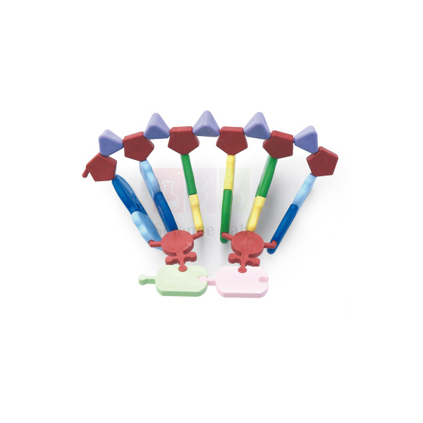
Molecular Model, RNA 12 Base
Molecular Model, RNA 12 Base (Product Code: SLE/Mo/20) is a molecular model kit used to build a 12-base section of RNA, the nucleic acid that carries genetic information from DNA to the protein-making machinery of the cell. It helps students compare RNA with DNA and understand codons, and its precision-moulded parts join firmly and store in a compartment case. Jain Scientific Equipments Pvt Ltd (ScienceLab), an OEM manufacturer, makes the RNA model at its own factory in Ambala, India.
Key Details
| Parameter | Detail |
|---|---|
| Model | RNA, 12 bases (as named) |
| Joining pieces | Designed for a firmly constructed model that can be passed around |
| Moulds and materials | Precision-engineered |
| Storage | Durable storage case with compartments for segregating parts |
| Part count and assembled size | Not stated in this listing — ask us |
| Product code | SLE/Mo/20 |
| Manufacturer | Jain Scientific Equipments Pvt Ltd (ScienceLab) — OEM |
| Country of origin | India (made in Ambala, Haryana) |
What Is the RNA Model Used For?
RNA differs from DNA in three ways that students need to learn: its sugar is ribose rather than deoxyribose, it uses the base uracil in place of thymine, and it is usually a single strand. A model built base by base makes these differences concrete, especially when placed beside a DNA model.
Twelve bases divide neatly into four codons of three bases each, so the model is well suited to reading a short message with a genetic code table and naming the amino acids it would specify. Teachers also use it to show transcription — building the RNA strand to complement a DNA template — before moving on to translation and protein synthesis.
OEM Manufacturer
The Molecular Model, RNA 12 Base is manufactured by Jain Scientific Equipments Private Limited, trading as ScienceLab, as the OEM (original equipment manufacturer). It is made at the company's own licensed factory at Saha, Ambala, Haryana, India, not sourced through a trader.
Schools, colleges, dealers and importers receive quotations directly from the manufacturer, which also handles repeat orders. The ScienceLab profile has more about the company.
Related Molecule Models
Items that connect the RNA model to DNA and protein synthesis:
- DNA-RNA Model — for side-by-side comparison of the two nucleic acids.
- Molecular Model Kit, Ten Layer DNA — a DNA helix to use as the template in transcription lessons.
- Protein Biosynthesis — follows the message from RNA to a finished protein.
- Molecular Model Set, 7 Amino Acid Collection — models of amino acids, the units that codons specify.
More nucleic acid models are listed in the Molecule range.
Frequently Asked Questions
What does "12 base" mean in the Molecular Model, RNA 12 Base?
The Molecular Model, RNA 12 Base builds an RNA section with 12 bases. That length divides into four three-base codons, which makes the model convenient for teaching how a genetic message is read and translated.
How is the RNA model different from the DNA model kits?
The Molecular Model, RNA 12 Base represents RNA, which uses ribose sugar and the base uracil and is usually single-stranded. The DNA kits in the Molecule range build double-stranded DNA, so using both highlights the differences between the two nucleic acids.
Will the RNA model hold together during class?
Yes, the Molecular Model, RNA 12 Base uses joining pieces designed so the model stays firmly constructed. The finished strand can be passed around the class during a lesson without falling apart.
Who manufactures the Molecular Model, RNA 12 Base?
The Molecular Model, RNA 12 Base is manufactured by Jain Scientific Equipments Pvt Ltd (ScienceLab) as the OEM at its own licensed factory in Saha, Ambala, Haryana, India. Enquiries are answered directly by the manufacturer.
What is in the RNA model case?
The Molecular Model, RNA 12 Base comes in a durable storage case with compartments to keep its parts segregated. The exact part count is not stated in this listing, so ask us through the contact page to confirm contents with your quotation.
Which topics does the RNA 12 base model support?
The Molecular Model, RNA 12 Base supports lessons on nucleic acid structure, transcription, codons and the genetic code, and the link between RNA and protein synthesis in senior-secondary and undergraduate biology.
Request a Quote
To request a quotation for the Molecular Model, RNA 12 Base, contact us via the contact page or add it to the enquiry cart. The manufacturer handles your enquiry directly.
Last updated: 24 September 2026
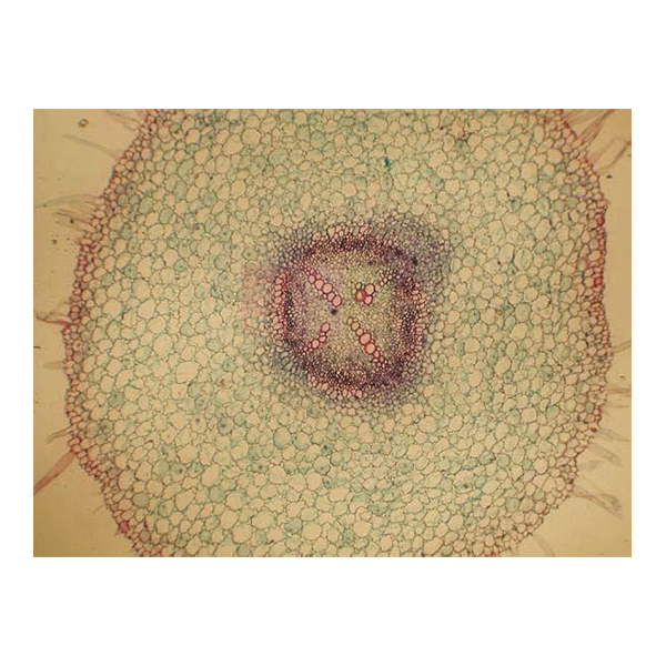
Basic Plant Anatomy Slide Set
Basic Plant Anatomy Slide Set (Product Code: SLE/PS/100) is a set of 20 glass microscope slides used to study the internal structure of vascular plants in school and college botany practicals. The prepared microscope slides cover roots, stems, leaves, flowers and seeds of plants such as Vicia, Zea, Lilium, Helianthus and Tilia, so one box supports a whole plant anatomy course. It is supplied by Jain Scientific Equipments Pvt Ltd (ScienceLab), Ambala, India.
Specifications
| Parameter | Detail |
|---|---|
| Slides in the set | 20 glass microscope slides |
| Theme | Major aspects of vascular plant structure |
| Roots | Vicia, root apex LS; Vicia (bean), young root TS; Helianthus (sunflower), young and old root TS |
| Stems | Zea (maize), stem TS and LS; Dicot, stem, apex LS; Cucurbita (marrow), stem TS and LS; Helianthus, stem, bundles TS; Helianthus, old stem TS and LS; Tilia (lime), young and old stem TS; Tilia, old stem RLS |
| Leaves and epidermis | Prunus (cherry), leaf TS; Ammophila (marram grass), leaf TS; Monocot, epidermis, for stomata E |
| Flowers and pollen | Lilium, anther TS; Lilium, ovary TS and LS; Lilium, ovary, various stages TS; Stigma with pollen tubes; Pollen grains |
| Seeds and embryo | Zea, seed LS; Capsella, mature cotyledon stage LS |
| Product code | SLE/PS/100 |
| Supplied by | Jain Scientific Equipments Pvt Ltd (ScienceLab), Ambala, India |
Label abbreviations: TS = transverse section, LS = longitudinal section, RLS = radial longitudinal section, E = entire (whole mount).
What Is the Basic Plant Anatomy Slide Set Used For?
Students use the set under a compound microscope to compare tissues across plant organs. Paired sections such as Zea and Cucurbita stems in both TS and LS show how vascular bundles look when cut across and along, while young and old stems of Tilia and Helianthus reveal secondary growth. The Vicia root apex and young root sections show the growing tip and the arrangement of root tissues.
For reproduction and adaptation topics, the Lilium anther and ovary slides, stigma with pollen tubes and pollen grains cover the flower, the Capsella slide shows an embryo at the mature cotyledon stage, and the marram grass leaf is a classic example of a leaf adapted to dry, windy sites. A slide box keeps the set in order, and the slides serve well as reference material in any botany lab equipment list for practical examinations.
What to Specify When Ordering
- Number of Basic Plant Anatomy Slide Sets (one per bench or one per class)
- Whether storage such as a slide box should be quoted with the sets
- Whether you also need microscopes for the practical
Supplier
Jain Scientific Equipments Private Limited supplies the Basic Plant Anatomy Slide Set under its ScienceLab trading name from Ambala, Haryana, serving schools, universities, dealers and importers in India and international markets. For tender and vendor forms, the supplying company's details are Importer-Exporter Code AAECJ8618A (DGFT) and GSTIN 06AAECJ8618A1ZT. See our company profile for more.
Related Prepared Slides
Useful additions for a plant anatomy practical:
- Prepared Microscope Slide Set – Transport In Plants — multiple copies of stem, root and leaf sections for a whole class
- Basic Botany Slide Set — covers algae, fungi, mosses and ferns alongside flowering plants
- Cell Division Slide Set of 9 — adds mitosis and meiosis to the plant course
- Slide Box — storage for the 20 slides
- Monocular Biological Microscope 400x, LED — a student microscope for viewing the sections
View all Prepared Slides.
Frequently Asked Questions
How many slides are in the Basic Plant Anatomy Slide Set?
The Basic Plant Anatomy Slide Set contains 20 glass microscope slides showing the major aspects of vascular plant structure, covering roots, stems, leaves, flowers, pollen, a seed and an embryo.
Which plants are used in the Basic Plant Anatomy Slide Set?
The Basic Plant Anatomy Slide Set uses Vicia (bean), Zea (maize), Lilium, Tilia (lime), Prunus (cherry), Cucurbita (marrow), Helianthus (sunflower), Capsella, Ammophila (marram grass) and a monocot epidermis prepared for viewing stomata.
What do TS, LS, RLS and E mean on the Basic Plant Anatomy Slide Set?
On the Basic Plant Anatomy Slide Set, TS means transverse section, LS longitudinal section, RLS radial longitudinal section and E an entire, or whole-mount, preparation. Several slides carry both TS and LS cuts of the same organ.
What microscope is needed for the Basic Plant Anatomy Slide Set?
The Basic Plant Anatomy Slide Set is viewed with an ordinary compound microscope of the kind used in school and college biology labs. Tell us if you need microscopes as well, and we will include them in the same quotation.
Who supplies the Basic Plant Anatomy Slide Set?
The Basic Plant Anatomy Slide Set is supplied by Jain Scientific Equipments Pvt Ltd (ScienceLab), Ambala, Haryana, India. The company provides IEC and GSTIN details to institutions and importers that need them for registration or export paperwork.
How is the Basic Plant Anatomy Slide Set different from the Basic Botany Slide Set?
The Basic Plant Anatomy Slide Set focuses on the internal structure of flowering and other vascular plants in 20 slides, whereas the Basic Botany Slide Set spreads its slides across the major plant groups, including algae, fungi, mosses and ferns.
Request a Quote
Ask for a quotation for the Basic Plant Anatomy Slide Set through our contact page, or add it to the enquiry cart with any microscopes or storage you need.
Last updated: 24 September 2026
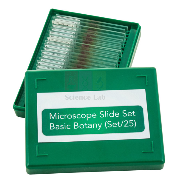
Basic Botany Slide Set
Basic Botany Slide Set (Product Code: SLE/PS/101) is a set of 25 glass microscope slides of botanical specimens used to survey the major plant groups in school and college botany classes. It runs from single-celled Chlamydomonas and Spirogyra through fungi, liverworts, mosses and ferns to pine and flowering plants, making it a ready collection of permanent slides for biology classification work. It is supplied by Jain Scientific Equipments Pvt Ltd (ScienceLab), Ambala, India.
Specifications
| Parameter | Detail |
|---|---|
| Quantity | 25 glass microscope slides |
| Algae | Chlamydomonas E; Spirogyra, vegetative E; Spirogyra, conjugation E; Fucus, dioecious antheridial and oogonial conceptacles |
| Fungi and yeast | Mucor, conjugation; Saccharomyces (yeast) budding; Penicillium, mycelium and conidia |
| Liverworts and mosses | Marchantia (liverwort) thallus, antheridia VS; Marchantia, thallus, archegonia VS; Marchantia, developing sporogonium VS; Mnium, archegonial head VS; Mnium (moss) antheridial head VS |
| Ferns | Dryopteris (fern), pinnule with sori TS; Dryopteris prothallus with antheridia and archegonia E |
| Conifer | Pinus (pine) leaf TS |
| Flowering plants | Zea (maize) root TS; Zea stem TS; Helianthus stem TS; Helianthus (sunflower) young root TS; Prunus (cherry) leaf TS; Tilia (lime) young and old stem TS; Cucurbita (marrow) stem, for sieve tubes LS; Ranunculus (buttercup) flower bud LS; Pollen grains, wind and insect distribution E; Allium (onion) root apex, mitosis LS |
| Product code | SLE/PS/101 |
| Supplied by | Jain Scientific Equipments Pvt Ltd (ScienceLab), Ambala, India |
What Is the Basic Botany Slide Set Used For?
The set is built for comparing groups rather than studying one plant in depth. A class can place the Marchantia, Mnium and Dryopteris slides side by side to see antheridia and archegonia in a liverwort, a moss and a fern, then contrast them with the Ranunculus flower bud and pollen grains adapted for wind and insect distribution. The algae and fungi slides show conjugation in Spirogyra and Mucor, budding yeast and Penicillium conidia.
The flowering-plant slides add everyday anatomy practice with root, stem and leaf sections, while the Cucurbita LS is chosen for sieve tubes and the Allium root apex for stages of mitosis. Teachers often keep the set as a reference collection in the botany lab and bring out the relevant slides as each plant group is taught.
Supplier
The Basic Botany Slide Set is supplied by Jain Scientific Equipments Private Limited, the Ambala, Haryana company that trades as ScienceLab. We source prepared slides for schools, colleges, laboratories and dealers in India and international markets and can quote them alongside microscopes and slide storage. Find out more on our company profile.
Related Prepared Slides
Other items used with a plant diversity course:
- Basic Plant Anatomy Slide Set — 20 slides for deeper study of vascular plant structure
- Cell Division Slide Set of 9 — builds on the Allium mitosis slide
- Basic Slide Set of 25 — a mixed plant and animal set for general biology
- Slide Box, Wooden — keeps the 25 slides organised
- Binocular Biological Microscope — for examining the specimens
Explore our Prepared Slides category for more sets.
Frequently Asked Questions
Which plant groups does the Basic Botany Slide Set cover?
The Basic Botany Slide Set covers algae, fungi and yeast, liverworts, mosses, ferns, a conifer (pine) and flowering plants, using 25 glass microscope slides that represent the major plant groups studied in botany.
Does the Basic Botany Slide Set show plant reproduction?
Yes. The Basic Botany Slide Set includes antheridia and archegonia of Marchantia, Mnium and a Dryopteris prothallus, a developing Marchantia sporogonium, fern sori, Fucus conceptacles, conjugation in Spirogyra and Mucor, a Ranunculus flower bud and pollen grains.
What do the letters E, VS, TS and LS mean on the Basic Botany Slide Set?
On the Basic Botany Slide Set, E marks an entire (whole-mount) specimen, VS a vertical section, TS a transverse section and LS a longitudinal section, telling students how each specimen was cut before mounting.
Is the Basic Botany Slide Set suitable for class practicals?
The Basic Botany Slide Set suits teacher demonstrations and small-group practicals because it holds one slide of each of 25 specimens. For whole-class work on a single topic, schools usually order extra sets or a multi-copy set.
Who supplies the Basic Botany Slide Set?
The Basic Botany Slide Set is supplied by Jain Scientific Equipments Pvt Ltd (ScienceLab), Ambala, Haryana, India, to schools, colleges and dealers in India and international markets.
What should I include in a quotation request for the Basic Botany Slide Set?
For the Basic Botany Slide Set, give product code SLE/PS/101, the number of sets, any microscopes or slide boxes required and the delivery address or country. Send them via our contact page for a prompt reply.
Request a Quote
Enquire about the Basic Botany Slide Set on our contact page, or add it to the enquiry cart with the other slides and equipment for your botany lab.
Last updated: 24 September 2026
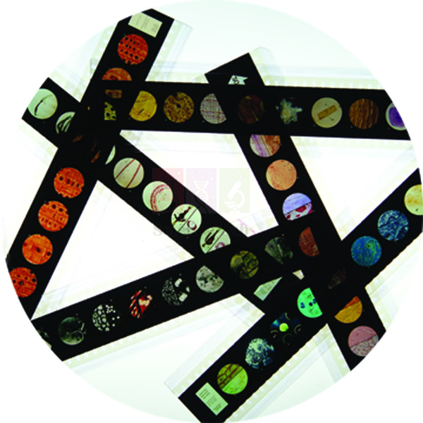
Microslide The Root Of a Flowering Plant
Microslide The Root Of a Flowering Plant (Product Code: SLE/PS/102) is a Microslide set of related 35 mm images photographed through a microscope, used to teach root structure and root growth in plant biology lessons. Its named views follow a buttercup root from the central cylinder, epidermis and cortex down through the root cap and the zones of growth, elongation and maturation to the root hairs. It is supplied by Jain Scientific Equipments Pvt Ltd (ScienceLab), Ambala, India.
Key Details
| Parameter | Detail |
|---|---|
| Format | Microslides are sets of 8 related 35 mm images photographed through a microscope |
| Root structure views | Buttercup Root; Central Cylinder; Epidermis and Cortex |
| Root growth views | Root Cap and Zone of Growth; Zone of Elongation; Zone of Maturation; Root Tip – Root hairs |
| Annotations | Arrows and callouts help students locate the features being studied |
| Text folder | Detailed folder to stimulate, inform and question students, with a pocket for storing the Microslides |
| Viewing device | Microslide Viewer |
| Product code | SLE/PS/102 |
| Supplied by | Jain Scientific Equipments Pvt Ltd (ScienceLab), Ambala, India |
What Is the Microslide The Root Of a Flowering Plant Used For?
The set helps students connect what a root looks like inside with how it grows. The buttercup root views show the outer epidermis, the wide cortex and the central cylinder that carries water and food, which is the standard dicot root pattern examined in school syllabuses.
The growth sequence then moves along the tip: the root cap protecting the zone of growth, cells lengthening in the zone of elongation, and tissues specialising in the zone of maturation, where root hairs increase the surface for taking up water. Because the callouts point to each zone, pupils can label a root-tip diagram straight from the Microslide Viewer before trying a root section under the microscope. It is a simple piece of plant biology lab equipment for revision and for classes without a slide-preparation facility.
Supplier
Jain Scientific Equipments Private Limited, which does business as ScienceLab in Ambala, Haryana, supplies Microslide The Root Of a Flowering Plant to schools, colleges and education suppliers. We handle orders for India and international markets and can add viewers or glass slide sets to the same quotation. Company background is available on our profile page.
Related Prepared Slides
Items that extend a lesson on roots:
- Microslide Viewer — needed to look at the images
- Basic Plant Anatomy Slide Set — glass slides including Vicia and sunflower root sections
- Microslide Photosynthesis — links root hairs and vascular tissue to leaf function
- Kit for Demonstrating Root Pressure — a practical on water uptake by roots
See every set in Prepared Slides.
Frequently Asked Questions
What views are named in Microslide The Root Of a Flowering Plant?
Microslide The Root Of a Flowering Plant names seven views: buttercup root, central cylinder, epidermis and cortex, root cap and zone of growth, zone of elongation, zone of maturation, and root tip with root hairs. Ask us for the complete image list.
How many images does Microslide The Root Of a Flowering Plant contain?
Microslides are described as sets of 8 related 35 mm images, while our listing for Microslide The Root Of a Flowering Plant names seven views. Please confirm the full contents with us through the contact page before ordering.
Which plant is shown in Microslide The Root Of a Flowering Plant?
Microslide The Root Of a Flowering Plant uses the buttercup root for its structural views, a commonly studied example of a flowering plant root, together with root-tip views of the growth zones and root hairs.
Is a text folder included with Microslide The Root Of a Flowering Plant?
Yes. Microslide The Root Of a Flowering Plant is accompanied by a detailed text folder that informs and questions students, and the folder has a handy pocket to keep the Microslides safe.
Who supplies Microslide The Root Of a Flowering Plant?
Microslide The Root Of a Flowering Plant is supplied by Jain Scientific Equipments Pvt Ltd (ScienceLab), Ambala, Haryana, India, which serves schools, colleges and dealers in India and international markets.
Request a Quote
To receive a quotation for Microslide The Root Of a Flowering Plant, use our contact page or add the set to the enquiry cart with any viewers you need.
Last updated: 24 September 2026
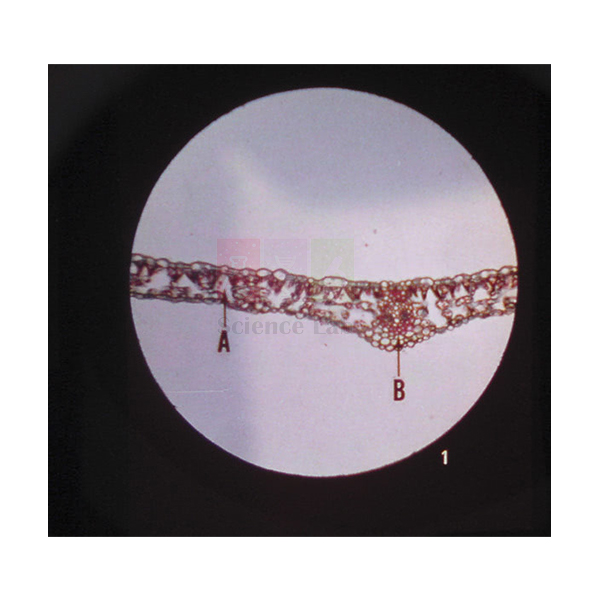
Microslide Photosynthesis
Microslide Photosynthesis (Product Code: SLE/PS/103) is a set of 8 related 35 mm images, photographed through a microscope, used to show where and how plants make and store food in school biology lessons on photosynthesis. It pairs green leaf structure at 110x and 350x with images of a leaf kept with and without sunlight, a variegated leaf, starch grains and a bean seed. The set is supplied by Jain Scientific Equipments Pvt Ltd (ScienceLab), Ambala, India.
Specifications
| Parameter | Detail |
|---|---|
| Contents | 8 related 35 mm images photographed through a microscope |
| Leaf structure | Green leaf (110x); Green leaf (350x) |
| Supply of raw materials | Root hair; Vascular bundles |
| Conditions for photosynthesis | Leaf with and without sunlight; Leaf – Variegated |
| Food storage | Starch grains; Bean seed |
| Student aids | Arrows and callouts on the images; detailed text folder with a pocket that holds the Microslides |
| Viewed in | Microslide Viewer |
| Product code | SLE/PS/103 |
| Supplied by | Jain Scientific Equipments Pvt Ltd (ScienceLab), Ambala, India |
What Is the Microslide Photosynthesis Used For?
Teachers use the set to turn photosynthesis from an equation into something students can see. The two green leaf images, at 110x and then 350x, let a class move from the overall layout of the leaf to a closer look at its cells. The root hair and vascular bundle frames show how water reaches the leaf.
The comparison of a leaf with and without sunlight and the variegated leaf image support the classic discussion of why light and chlorophyll are needed, and the starch grains and bean seed show where the products end up in storage. Students can study the callouts in a Microslide Viewer before or after running a starch test with a photosynthesis experiment kit, using the text folder questions to explain their results.
Supplier
Microslide Photosynthesis is sourced and supplied by Jain Scientific Equipments Private Limited, whose trading name is ScienceLab, from Ambala in Haryana, India. Our customers include schools, colleges and dealers in India and international markets who want slides, apparatus and kits quoted together. Read about the company on our company profile.
Related Prepared Slides
Pair the set with these photosynthesis resources:
- Microslide Viewer — for viewing the eight images
- Prepared Microscope Slide Set – Photosynthesis and Adaptations — glass slides for microscope practicals on leaves
- Photosynthesis Apparatus — demonstrates the process in a working experiment
- Kit for Photosynthesis Experiments — hands-on tests to match the images
- Microslide The Root Of a Flowering Plant — more detail on roots and root hairs
Browse more in Prepared Slides.
Frequently Asked Questions
What images are in Microslide Photosynthesis?
Microslide Photosynthesis contains green leaf at 110x, green leaf at 350x, root hair, vascular bundles, a leaf with and without sunlight, a variegated leaf, starch grains and a bean seed, all photographed through a microscope.
Why does Microslide Photosynthesis include a variegated leaf?
Microslide Photosynthesis includes a variegated leaf because its green and pale areas let students discuss the role of chlorophyll, alongside the leaf shown with and without sunlight, which relates to the need for light.
Can Microslide Photosynthesis be used without a microscope?
Yes. Microslide Photosynthesis is viewed in a Microslide Viewer rather than a microscope, because its images were already photographed through a microscope. Order viewers with the set if your school does not have them.
What written material comes with Microslide Photosynthesis?
Microslide Photosynthesis comes with a detailed text folder designed to stimulate, inform and question students, with a handy pocket where the Microslides are kept between lessons.
Who supplies Microslide Photosynthesis?
Microslide Photosynthesis is supplied by Jain Scientific Equipments Pvt Ltd (ScienceLab), Ambala, Haryana, India. The company quotes it for schools, colleges and dealers in India and international markets, together with related apparatus if needed.
What details should a Microslide Photosynthesis enquiry include?
A Microslide Photosynthesis enquiry should give code SLE/PS/103, the number of sets, whether viewers or photosynthesis apparatus are wanted, and the delivery location. Our contact page is the quickest way to send them.
Request a Quote
Request pricing for Microslide Photosynthesis via our contact page, or add it to the enquiry cart alongside other photosynthesis resources.
Last updated: 24 September 2026
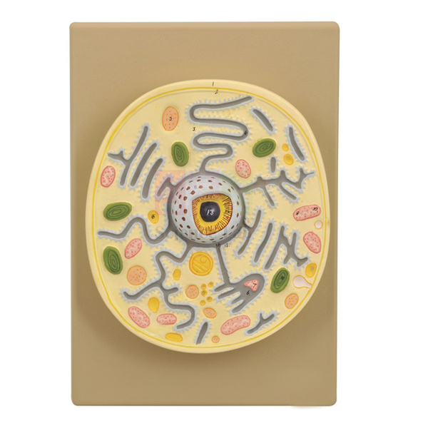
Plant Cell on Base Model
Plant Cell on Base Model (Product Code: SLE/ECB/20) is a base-mounted model of a plant cell's electron microscopic structure, used to teach cell ultrastructure in senior secondary, undergraduate botany and teacher-training classes. A portion of the wall is removed to reveal the ectoplast, endoplast and tonoplast, the vacuoles, nuclear structure, plastids and mitochondria. It is made by Jain Scientific Equipments Pvt Ltd (ScienceLab), an OEM manufacturer, at its own factory in Ambala, India, as an advanced plant anatomy model for institutions in India and abroad.
Key Details
| Parameter | Detail |
|---|---|
| Type | Plant cell model showing electron microscopic structure |
| Presentation | Portion of the cell wall removed to expose the interior |
| Structures shown | Ectoplast, endoplast, tonoplast, vacuoles, nuclear structure, plastids, mitochondria and other cell components |
| Mounting | On a base |
| Product code | SLE/ECB/20 |
| Manufacturer | Jain Scientific Equipments Pvt Ltd (ScienceLab) — OEM |
| Country of origin | India (made in Ambala, Haryana) |
What Is the Plant Cell on Base Model Used For?
The model is most useful once students already know the basic parts of a plant cell and are ready to study membranes in detail. With the wall partly cut away, a teacher can show that the cell contents are bounded by the ectoplast, that the vacuoles are enclosed by their own membrane, the tonoplast, and that the nucleus, plastids and mitochondria sit within the surrounding cytoplasm. This makes it easier to explain plasmolysis, where the protoplast pulls away from the wall, and how substances are stored in or released from the vacuole.
Because it depicts electron microscope detail, the model also helps students interpret published electron micrographs, which can otherwise look like abstract patterns. Colleges often display it beside a conventional plant cell model so that learners see the same cell at two levels of magnification, and the base keeps it steady on a bench or display shelf between lessons.
OEM Manufacturer
ScienceLab is the name used by Jain Scientific Equipments Private Limited, the OEM (original equipment manufacturer) of the Plant Cell on Base Model, which the company makes at its own licensed factory at Saha, Ambala, Haryana, India. Colleges, schools, dealers and importers therefore buy direct from the maker and can return to it for repeat orders or questions about the model — ask us. Buyers who need vendor identifiers for procurement can use the company's GST registration, 06AAECJ8618A1ZT, and its Legal Entity Identifier, 3358004HRD1JIVXKYX49. See the company profile for more.
Related Cell Biology Equipments
Models that complement an ultrastructure lesson on plant cells:
- Plant Cell — a larger translucent model with a multi-layered cell wall and 25 organelles and inclusions.
- Model Plant Cell — a compact model for group work at an introductory level.
- Chloroplast Model — an enlarged plastid for studying its internal membranes.
- Mitochondrion Model — enlarges another organelle shown on this model.
- Plant Cell Plaque — a plaque-format plant cell for comparison and revision.
Browse the full Cell Biology Equipments range.
Frequently Asked Questions
How is the Plant Cell on Base Model different from a standard plant cell model?
The Plant Cell on Base Model shows the electron microscopic structure of the cell, with part of the wall removed, so membranes such as the ectoplast and tonoplast can be studied along with the vacuoles, nuclear structure, plastids and mitochondria.
Is the Plant Cell on Base Model mounted?
Yes. The Plant Cell on Base Model is mounted on a base, which keeps it stable on a demonstration bench or display shelf and lets it stay out for the whole of a practical session without being handled.
What are the dimensions and weight of the Plant Cell on Base Model?
The size and weight of the Plant Cell on Base Model are not given in our current product information. Please ask through our contact page for dimensions and packed weight before ordering, especially for export shipments.
Which level of student is the Plant Cell on Base Model for?
The Plant Cell on Base Model is most useful from senior secondary level upward, where plant cell ultrastructure and membranes are part of the syllabus, and in undergraduate botany and teacher-training courses.
How should the Plant Cell on Base Model be cared for?
Dust the Plant Cell on Base Model with a soft brush or cloth, lift it by the base rather than the cut-away section, and keep it away from heat and direct sunlight so the surface and colours stay in good condition.
Who manufactures the Plant Cell on Base Model?
The Plant Cell on Base Model is manufactured by Jain Scientific Equipments Pvt Ltd (ScienceLab), the OEM, at its own licensed factory at Saha, Ambala, Haryana, India. Institutions and distributors order it and receive quotations directly from the manufacturer.
What should a quotation request for the Plant Cell on Base Model include?
A quotation request for the Plant Cell on Base Model should include product code SLE/ECB/20, the quantity, the delivery location and any tender format you need. Buyers in India and international markets are supplied.
Request a Quote
To get a quotation for the Plant Cell on Base Model, reach our team or add it to the enquiry cart; the quotation comes straight from the manufacturer.
Last updated: 24 September 2026
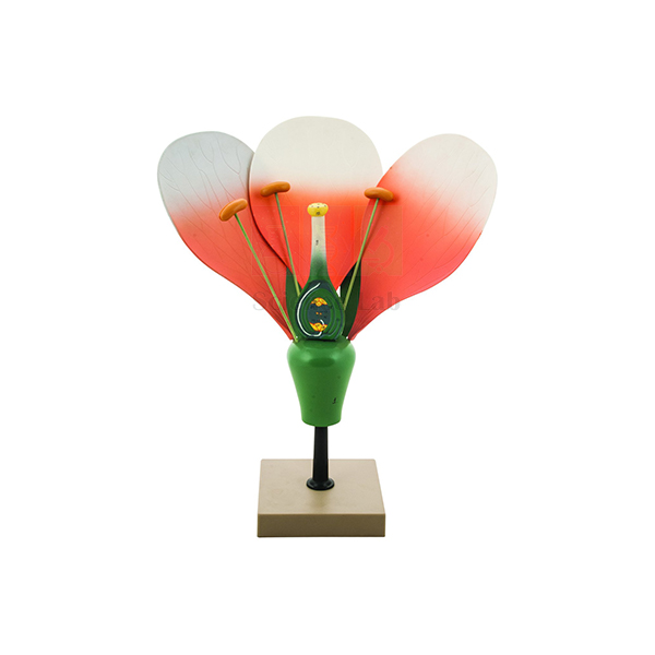
Typical Flower Model
Typical Flower Model (Product Code: SLE/EBT/17) is a large size teaching model used for detailed study of the structure and functional parts of a flower in school and college biology classes. Every part is removable for better viewing and the ovary is bisected to reveal internal structures, so teachers can dismantle the flower whorl by whorl as a hands-on plant anatomy model. It is made by Jain Scientific Equipments Pvt Ltd (ScienceLab), an OEM manufacturer, at its own factory in Ambala, India.
Key Details
| Parameter | Detail |
|---|---|
| Type | Large size model of a typical flower |
| Purpose | Detailed study of the structure and functional parts of a flower |
| Removable parts | All parts removable for better viewing |
| Ovary | Bisected to show internal structures |
| Product code | SLE/EBT/17 |
| Manufacturer | Jain Scientific Equipments Pvt Ltd (ScienceLab) — OEM |
| Country of origin | India (made in Ambala, Haryana) |
What Is the Typical Flower Model Used For?
Teachers usually take the model apart in the same order a botanist would dissect a real flower: first the outer whorls that protect and attract, then the male parts, and finally the female parts at the centre. Lifting each removable piece away lets the class see where it attaches and how it relates to its neighbours, which is much harder to show with a drawing. The bisected ovary then opens the discussion on ovules, fertilisation and how the ovary develops into a fruit after pollination.
After the demonstration, students can reassemble the model themselves as a quick check of their understanding, or use it as a reference while dissecting a real flower with forceps and a hand lens. Among biology teaching aids in school labs across India, a removable-part flower model is a practical way to prepare students for labelled-diagram questions. Count the parts at the end of each lesson and store the model assembled so no pieces are lost.
OEM Manufacturer
Jain Scientific Equipments Private Limited, known by its trading name ScienceLab, is the OEM (original equipment manufacturer) of the Typical Flower Model, which is produced at the company's own licensed factory at Saha, Ambala, Haryana. Schools, colleges, dealers and importers order it direct from the maker, and questions about a missing piece or a repeat order are handled by the manufacturer itself — just ask us. See the company profile for more.
Related Botany Equipment
Items used with this model in lessons on flower structure and reproduction:
- Typical Flower V.S. Model — a vertical-section model on a base, showing the flower cut through its centre.
- Plant Dicot Flower — for comparing the general plan with a dicot flower.
- Flower Formation in Angiosperms — extends the lesson to how flowers develop.
- Dissecting Set, Student — tools for dissecting real flowers after the model demonstration.
- Magnifying Glass 2 — for inspecting pollen, anthers and stigmas on fresh flowers.
For more flower and plant models, browse the Botany category.
Frequently Asked Questions
Which parts of the Typical Flower Model can be removed?
All parts of the Typical Flower Model are removable for better viewing, so a teacher can take the flower apart during a lesson and students can rebuild it afterwards as a quick check of what they have learned.
What does the bisected ovary show?
The bisected ovary of the Typical Flower Model opens to reveal its internal structures, which helps a teacher explain where ovules sit and how fertilisation leads to seed and fruit formation after pollination.
What are the exact dimensions?
The Typical Flower Model is described as large size, but exact dimensions are not stated in our listing. Ask through our contact page with code SLE/EBT/17 and ScienceLab will confirm the measurements and packed size.
How does it differ from the Typical Flower V.S. Model?
The Typical Flower Model has removable parts and a bisected ovary for hands-on dismantling, whereas the Typical Flower V.S. Model shows a fixed vertical section mounted on a base. Many departments use both side by side.
Who manufactures the Typical Flower Model?
The Typical Flower Model is manufactured by Jain Scientific Equipments Pvt Ltd (ScienceLab), the OEM, at its own licensed factory in Saha, Ambala, Haryana, India, so buyers deal with the maker directly for quotations and follow-up questions.
Can a lost part be replaced?
If a piece of the Typical Flower Model goes missing, contact ScienceLab with code SLE/EBT/17 and a description of the part; as the manufacturer, the company will advise whether a replacement can be supplied.
Can you supply several models to one institution?
Yes. ScienceLab supplies the Typical Flower Model in quantity to schools, colleges and distributors in India and international markets; include the number of models and delivery location in your enquiry for a factory-direct quotation.
Request a Quote
Request a price for the Typical Flower Model on our contact page, or add it to the enquiry cart together with the V.S. model and dissecting tools for a complete flower lesson; quotations come straight from the manufacturer.
Last updated: 24 September 2026
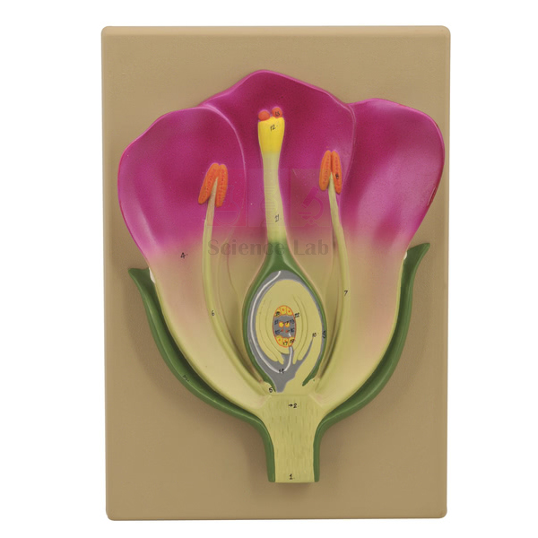
Typical Flower V.S. Model
Typical Flower V.S. Model (Product Code: SLE/EBT/18) is a vertical-section model of a flower used to show how every floral part is arranged, from receptacle to stigma, in school and college botany lessons. Mounted on a base and weighing 650 g, it stands securely for demonstrations yet is light enough to pass along a bench. It is made by Jain Scientific Equipments Pvt Ltd (ScienceLab), an OEM manufacturer, at its own factory in Ambala, India, as part of its botany lab equipment range.
Specifications
| Parameter | Detail |
|---|---|
| Type | Vertical section (V.S.) model of a flower |
| Mounting | Mounted on base |
| Weight | 650 g |
| Dimensions | 330 x 250 x 50 (as listed; unit not stated) |
| Product code | SLE/EBT/18 |
| Manufacturer | Jain Scientific Equipments Pvt Ltd (ScienceLab) — OEM |
| Country of origin | India (made in Ambala, Haryana) |
What Is the Flower V.S. Model Used For?
A vertical section is the view most textbooks and examinations use for flower structure, because it shows at a glance how the sepals, petals, stamens and carpel are attached to the receptacle and where the ovary sits in relation to the other parts. With the model on the bench, a teacher can trace the route a pollen grain takes from the stigma down the style to an ovule, and students can copy a labelled V.S. drawing from a three-dimensional example rather than from another diagram.
It is especially useful just before students cut their own vertical sections of fresh flowers with a scalpel, since they know what they are aiming to see. As one of the everyday biology teaching aids on a demonstration table or display shelf, it stays upright on its base, and its modest weight makes it easy for groups to share. Dust it regularly with a soft brush and avoid placing it where it may be knocked off a high shelf.
OEM Manufacturer
ScienceLab, the trading name of Jain Scientific Equipments Private Limited, is the OEM (original equipment manufacturer) of this sectional flower model and produces it at its own licensed factory in Saha, Ambala, Haryana. The company sells its biology, chemistry and physics models straight from the factory to schools, colleges, dealers and importers, and repeat orders are arranged with the maker rather than a trading intermediary. The company profile has more.
Related Botany Equipment
Complementary items for teaching flower structure:
- Typical Flower Model — a large model with removable parts to accompany the sectional view.
- Dicotyledonous Flower Pattern — for relating the general plan to dicot flowers.
- Monocotyledonous Flower Pattern — for comparing with monocot flower arrangement.
- Hand Lens — for examining students' own flower sections after the demonstration.
See the Botany category for further flower and plant anatomy models.
Frequently Asked Questions
What does V.S. stand for?
V.S. stands for vertical section: the Typical Flower V.S. Model shows a flower as if cut lengthwise down its centre, revealing how sepals, petals, stamens and carpel are attached and where the ovary sits.
How much does the Typical Flower V.S. Model weigh?
The Typical Flower V.S. Model weighs 650 g, so it is easy to carry between classrooms and to pass along a bench, while its base keeps it stable during a demonstration.
What are the dimensions?
The listed dimensions of the Typical Flower V.S. Model are 330 x 250 x 50. The unit is not stated in our listing, so ask through our contact page to confirm the measurements and the packed carton size.
Does it have removable parts?
Removable parts are not listed for the Typical Flower V.S. Model, which shows a fixed sectional view. If you need a flower that can be taken apart, the Typical Flower Model with removable parts is listed alongside it.
Who manufactures the Typical Flower V.S. Model?
The Typical Flower V.S. Model is manufactured by Jain Scientific Equipments Pvt Ltd (ScienceLab), the OEM, at its own licensed factory in Saha, Ambala, Haryana, India, and is supplied factory-direct to schools, colleges, dealers and importers.
How should the model be cleaned?
Clean the Typical Flower V.S. Model with a soft brush or dry cloth, avoid solvents, and never immerse it in water. Keep it on a low, stable shelf where it cannot be knocked off.
What details should an institutional order include?
An order for the Typical Flower V.S. Model should give code SLE/EBT/18, quantity, billing entity and delivery address; ScienceLab then quotes directly as the manufacturer for buyers in India and international markets.
Request a Quote
Ask for a manufacturer's quotation on the Typical Flower V.S. Model through the contact page, or place it in the enquiry cart with any other botany models you plan to buy.
Last updated: 24 September 2026