- Physics Lab Equipment
- Chemistry Lab Equipment
- Biology Lab Equipment
- Anatomical Lab Equipment
- Learning
- Educational Lab Equipment
- Magnets
- Agriculture
- Astronomy
- Earth Science
- Geography Lab Equipment
- Safety Lab Coat
- Laboratory
- Meteorological
- Electricity & Magnetism
- Heat
- School Lab Equipment
- Lab Glassware
- Science Lab Equipment
- Tvet Lab Equipment
- Robotics Kits Supplier and Manufacturer for Schools India
- Math Lab Equipment Manufacturers and Exporters in Ambala
- STEM Kits Manufacturers and Supplier India
- Engineering Lab Equipment
- Hospital Lab Equipment
- Water, Sanitation & Hygiene Supplies
- Cold Chain, Solar & Communication Equipment
- Nutrition
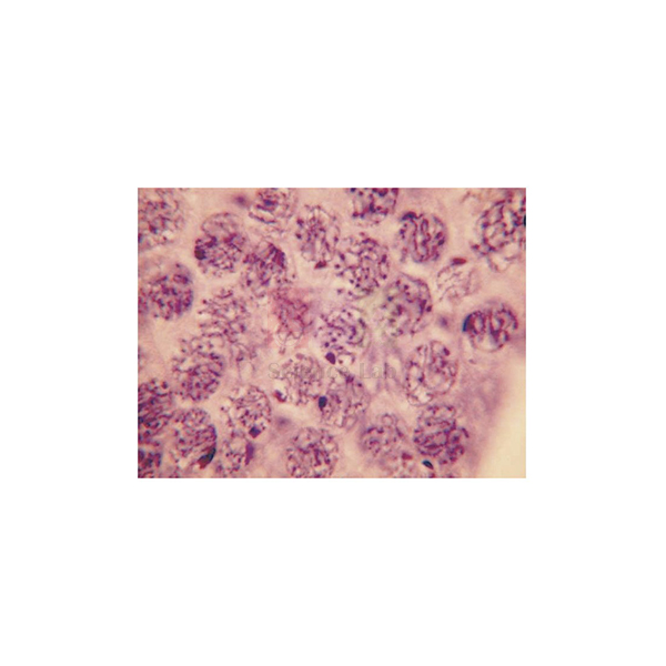
Cell Division Slide Set of 9
Cell Division Slide Set of 9 (Product Code: SLE/PS/80) is a set of nine prepared glass microscope slides used to show how chromosomes divide in plant and animal cells in school and college biology practicals. Teachers pick it because one box covers mitosis in Allium root tips, meiosis in Lilium and locust tissue, and an acetic-orcein squash of giant salivary-gland chromosomes. These prepared microscope slides are supplied by Jain Scientific Equipments Pvt Ltd (ScienceLab), Ambala, India.
Specifications
| Parameter | Detail |
|---|---|
| Slide type | Prepared glass microscope slides |
| Number of slides | 9 (listed below) |
| Topic | Chromosome division in animal and plant cells (mitosis and meiosis) |
| Plant material | Allium root apex and root tip; Lilium ovary and anther |
| Animal material | Locusta testis; Rana ova; insect salivary gland; mammal testis |
| Stain named in the set list | Acetic orcein (insect salivary gland squash) |
| Product code | SLE/PS/80 |
| Supplied by | Jain Scientific Equipments Pvt Ltd (ScienceLab), Ambala, India |
Slides in the set:
- Allium root apex LS, mitosis
- Allium root tip squash preparation
- Locusta testis VS
- Rana ova, various stages
- Lilium ovary, meiosis
- Lilium anther TS, three stages of development
- Insect salivary gland squash, acetic orcein, for giant chromosomes
- Mammal testis TS showing spermatogenesis
- Locusta testis squash showing meiosis
What Is the Cell Division Slide Set Used For?
A typical mitosis practical starts with the Allium root apex LS, where students scan the meristem just behind the root cap and count cells in prophase, metaphase, anaphase and telophase. The root tip squash then shows the same stages with the cells spread flat, which makes chromosome counting easier. For meiosis, the Lilium ovary and the three-stage Lilium anther section are compared with the locust testis squash, and the mammal testis TS links meiosis to sperm formation.
The Rana ova slide shows eggs at various stages, and the acetic-orcein salivary gland squash introduces giant chromosomes with visible banding, a useful bridge into genetics. As a microscope slide set for school and college labs, it gives the clearest results when students start on low power to locate the tissue and then move to higher power for chromosome detail.
Supplier
Jain Scientific Equipments Private Limited trades under the name ScienceLab and supplies this cell division set together with the microscopes, slide boxes and other prepared slide collections that a biology department orders for its practical course. The company works from Ambala, Haryana, and serves schools, colleges, training institutes and dealers in India and international markets. Background on the company is on the ScienceLab company profile page.
Related Prepared Slides
These items are often ordered with the Cell Division Slide Set of 9 for the same cell-division lessons:
- Mitosis & Meiosis Set, Microscope Slides — a second slide set on the two division types for larger practical groups.
- Compound Microscope — needed to view the root-tip and squash preparations at higher power.
- Slide Box — keeps the nine slides flat, labelled and protected between classes.
- Microslide Meiosis — photographic images of meiosis stages for pre-lab briefing or revision.
- Human Histology Slide Set of 25 — the next step once students move from cell division to tissues.
See the full range of Prepared Slides for other botany, zoology and histology sets.
Frequently Asked Questions
Which slides are included in the Cell Division Slide Set of 9?
The Cell Division Slide Set of 9 contains Allium root apex LS, Allium root tip squash, Locusta testis VS, Locusta testis squash, Rana ova at various stages, Lilium ovary, Lilium anther TS in three stages, an insect salivary gland squash and a mammal testis TS.
Does the Cell Division Slide Set of 9 cover both mitosis and meiosis?
Yes. The Cell Division Slide Set of 9 shows mitosis in the two Allium root preparations and meiosis in the Lilium ovary, the Lilium anther stages and the locust testis squash, while the mammal testis TS shows spermatogenesis.
What microscope is needed for the Cell Division Slide Set of 9?
Any compound microscope with low- and high-power objectives can be used with the Cell Division Slide Set of 9. Locate the dividing region on low power first, then switch to high power to study chromosome shape and arrangement in the squash preparations.
How should the Cell Division Slide Set of 9 be stored?
Store the Cell Division Slide Set of 9 flat in a slide box, away from heat, damp and direct sunlight. Hold slides by the edges, and wipe off immersion oil or fingerprints with lens tissue after each class so the preparations stay clear.
Who supplies the Cell Division Slide Set of 9?
The Cell Division Slide Set of 9 is supplied by Jain Scientific Equipments Pvt Ltd (ScienceLab), Ambala, Haryana, India. The company supplies prepared slides to schools, colleges, institutions and dealers in India and international markets.
What should a quotation request for the Cell Division Slide Set of 9 include?
For a quotation on the Cell Division Slide Set of 9, state the number of sets, the delivery city or country and any microscopes or slide boxes you need with it. Send these details through the contact page.
Request a Quote
To order the Cell Division Slide Set of 9 for your biology lab, send your quantity and delivery details through the contact page, or add it to the enquiry cart together with the other slides and microscopes on your list.
Last updated: 24 September 2026
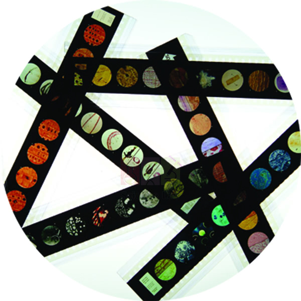
Microslide Animal Tissue Set 2 Muscle, Bone, Connective
Microslide Animal Tissue Set 2 Muscle, Bone, Connective (Product Code: SLE/PS/81) is a Microslide of 8 related 35mm images, photographed through a microscope, used to teach muscle, bone and connective tissue in school biology lessons. Students compare voluntary, involuntary and heart muscle alongside areolar tissue, bone, blood, fat and spinal cord, with arrows and callouts marking each key feature. This biology teaching aid is supplied by Jain Scientific Equipments Pvt Ltd (ScienceLab), Ambala, India.
Specifications
| Parameter | Detail |
|---|---|
| Format | Microslide: 8 related 35mm images photographed through a microscope |
| Viewed with | Microslide Viewer |
| Muscle images | Voluntary muscle cells, involuntary muscle cells, heart muscle |
| Connective and supporting tissue images | Areolar connective tissue, bone, blood, adipose tissue |
| Nervous tissue image | Spinal cord |
| Labelling | Arrows and callouts on the images |
| Accompanying material | Detailed text folder with a pocket for storing the Microslide |
| Product code | SLE/PS/81 |
| Supplied by | Jain Scientific Equipments Pvt Ltd (ScienceLab), Ambala, India |
What Is the Microslide Animal Tissue Set Used For?
The three muscle images set up the classic comparison in any tissue topic: long striated voluntary fibres, spindle-shaped involuntary cells and the branched fibres of heart muscle. Connective tissue then appears in four very different forms, from loose areolar tissue and fat-storing adipose tissue to the hard matrix of bone and the fluid tissue of blood. The spinal cord image rounds off the set with nervous tissue.
Because every image already carries arrows and callouts, teachers use this Microslide for a short briefing before students examine their own tissue slides, or for revision where microscope time is limited. The questions in the text folder work as a ready worksheet, which makes the set a practical biology teaching aid for large classes.
Supplier
ScienceLab is the trading name of Jain Scientific Equipments Private Limited, based in Ambala, Haryana, which supplies Microslide sets, viewers and prepared slides to schools, colleges and dealers in India and international markets. For vendor registration, the supplying company is listed under CIN U29309HR2020PTC087597 and GSTIN 06AAECJ8618A1ZT. More about the company is on the ScienceLab profile page.
Related Prepared Slides
Items that support the same tissue lessons as this Microslide:
- Microslide Viewer — the viewer these 35mm images are designed for.
- Microslide Basic Human Histology — adds cartilage, epithelium and nerve cells to the tissue unit.
- Microslide Cells of Body — looks at individual body cells rather than whole tissues.
- Prepared Microscope Slide Set – Connective Tissues — glass slides for students to examine under a microscope.
Browse all Prepared Slides for further Microslide topics and glass slide sets.
Frequently Asked Questions
Which images are in the Microslide Animal Tissue Set 2?
The Microslide Animal Tissue Set 2 Muscle, Bone, Connective has eight images: voluntary muscle cells, involuntary muscle cells, heart muscle, areolar connective tissue, bone, blood, adipose tissue and spinal cord, each marked with arrows and callouts.
Is the Microslide Animal Tissue Set 2 a glass microscope slide?
No. The Microslide Animal Tissue Set 2 is a set of 8 related 35mm images photographed through a microscope and viewed through a Microslide Viewer. For glass slides of similar tissues, see the connective tissues prepared slide set.
What comes with the Microslide Animal Tissue Set 2?
The Microslide Animal Tissue Set 2 comes with a detailed text folder written to inform students and question them on the topic. The folder has a pocket that holds the Microslide, so images and notes stay together.
Which muscle types does the Microslide Animal Tissue Set 2 show?
The Microslide Animal Tissue Set 2 shows all three muscle types: voluntary (skeletal) muscle cells, involuntary (smooth) muscle cells and heart muscle, so students can compare fibre shape and arrangement on one set.
Who supplies the Microslide Animal Tissue Set 2?
The Microslide Animal Tissue Set 2 Muscle, Bone, Connective is supplied by Jain Scientific Equipments Pvt Ltd (ScienceLab), Ambala, Haryana, India, to schools, colleges and dealers in India and international markets.
What information is needed to quote for the Microslide Animal Tissue Set 2?
To quote for the Microslide Animal Tissue Set 2, we need the number of sets, whether Microslide Viewers are also required, and the delivery location. Send these through the contact page.
Request a Quote
Ask for a quotation on the Microslide Animal Tissue Set 2 Muscle, Bone, Connective through the contact page, or place it in the enquiry cart with any viewers and other Microslide topics you need.
Last updated: 24 September 2026
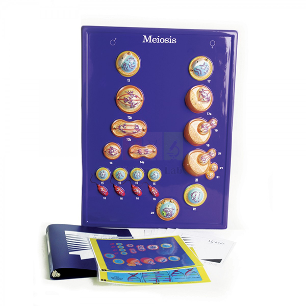
Meiosis Model
Meiosis Model (Product Code: SLE/PS/82) is an enlarged teaching model used to explain meiotic cell division in school and college cell biology lessons. Teachers use it to show how meiosis gives rise to individual human characteristics and genetic differences, with enlarged views of chromosomes, cytoplasm and the chromatic and polar bodies. It is made by Jain Scientific Equipments Pvt Ltd (ScienceLab), an OEM manufacturer, at its own factory in Ambala, India.
Key Details
| Parameter | Detail |
|---|---|
| Model type | Enlarged teaching model of meiotic cell division |
| Structures shown enlarged | Chromosomes, cytoplasm, chromatic bodies and polar bodies |
| Teaching focus | How meiosis leads to individual human characteristics and genetic differences |
| Subject area | Cell biology and genetics |
| Product code | SLE/PS/82 |
| Manufacturer | Jain Scientific Equipments Pvt Ltd (ScienceLab) — OEM |
| Country of origin | India (made in Ambala, Haryana) |
What Is the Meiosis Model Used For?
Meiosis is hard to picture from a flat textbook diagram, because chromosomes pair up, exchange segments and then separate in two rounds of division. The Meiosis Model enlarges those structures so a whole class can follow the sequence together, from the chromosomes in the cytoplasm to the appearance of polar bodies. It gives teachers a concrete way to answer the question students always ask: why do brothers and sisters look different?
Schools that buy a mitosis and meiosis model usually use it twice: at the start of a genetics unit and again when students compare the two kinds of division. During a slide practical, keep it on the front bench so students can match what they see under the microscope with its enlarged form. Dust it with a soft, dry cloth and store it away from direct sunlight.
OEM Manufacturer
ScienceLab is the trading name of Jain Scientific Equipments Private Limited, the OEM (original equipment manufacturer) of this Meiosis Model. The company makes its cell biology models at its own licensed factory at Saha, Ambala, Haryana, India, and sells them factory-direct to schools, colleges, institutions, dealers and importers instead of through a trading middleman.
Because the maker handles your order itself, repeat orders and questions about replacement parts go straight to the people who build the model — just ask us. Read more about the company on the ScienceLab profile page.
Related Cell Biology Equipments
These items are often used with the Meiosis Model in cell division and genetics lessons:
- Animal Mitosis Models — the matching models for comparing mitosis with meiosis.
- Animal Meiosis Plaque — a plaque presentation of meiosis in animal cells for wall or bench display.
- Mitosis Simulation Lab Investigation — a hands-on activity where students model division themselves.
- Mitosis & Meiosis Set, Microscope Slides — real dividing cells to view after studying the model.
- Microslide Meiosis — photographed stages of meiosis in the egg cell for revision.
See the complete Cell Biology Equipments range for cell models, plaques and investigations.
Frequently Asked Questions
What does the Meiosis Model show?
The Meiosis Model shows meiotic cell division with enlarged views of chromosomes, cytoplasm and the chromatic and polar bodies. It is designed to help students understand how meiosis produces individual human characteristics and genetic differences.
Who manufactures the Meiosis Model?
The Meiosis Model is manufactured by Jain Scientific Equipments Pvt Ltd (ScienceLab), the OEM, at its own licensed factory at Saha, Ambala, Haryana, India. Quotations and repeat orders come directly from the manufacturer.
Which classes is the Meiosis Model suitable for?
The Meiosis Model suits secondary school biology, where meiosis and inheritance are first introduced, and college cell biology or genetics courses that need a clear demonstration piece for lectures and practical sessions.
Can the Meiosis Model be used with a mitosis model?
Yes. The Meiosis Model is often ordered with the Animal Mitosis Models so teachers can place both divisions side by side and show how the number of chromosomes and the resulting cells differ between them.
Are dimensions and materials of the Meiosis Model available?
Dimensions and materials of the Meiosis Model are not listed on this page. Ask for them through the contact page and they will be confirmed with your quotation.
Can the Meiosis Model be ordered in bulk or for export?
Yes. The Meiosis Model is supplied factory-direct to schools, institutions, dealers and importers in India and international markets. Share the quantity and destination so the manufacturer can prepare a quotation.
Request a Quote
Quotations for the Meiosis Model come straight from the manufacturer. Send your requirement through the contact page, or add the model to the enquiry cart with the rest of your cell biology list.
Last updated: 24 September 2026
220.jpg)
Microslide Basic Human Histology
Microslide Basic Human Histology (Product Code: SLE/PS/83) is a Microslide of 8 related 35mm photomicrographs used to introduce the basic tissues of the human body in school and early college biology. Its images run from a stained cross-section of bone and cartilage to ciliated epithelium, stained nerve cells, striated muscle and red and white blood cells, each marked with arrows and callouts. It is supplied by Jain Scientific Equipments Pvt Ltd (ScienceLab), Ambala, India.
Specifications
| Parameter | Detail |
|---|---|
| Format | 8 related 35mm images photographed through a microscope |
| Viewing device | Microslide Viewer |
| Supporting tissue images | Bone XS (stained), cartilage, fat cells |
| Epithelial image | Epithelial tissue / ciliated epithelial cell |
| Nerve and muscle images | Nerve cells (stained), striated muscle cells |
| Blood images | Red blood cells, white blood cells |
| Student notes | Detailed text folder with questions, plus a pocket that stores the Microslide |
| Product code | SLE/PS/83 |
| Supplied by | Jain Scientific Equipments Pvt Ltd (ScienceLab), Ambala, India |
What Is the Microslide Basic Human Histology Used For?
Most histology courses begin with four tissue families: epithelial, connective, muscle and nervous. This Microslide covers all four, so a teacher can move from the cilia on epithelial cells to the stained nerve cells, the banding of striated muscle and the supporting matrix of bone and cartilage. The two blood images let students compare red and white cells before they attempt a blood smear of their own.
Students can keep the images beside them while drawing, using the callouts to label their diagrams correctly. Because it needs only a Microslide Viewer rather than a microscope and stains, the set is an easy addition to school biology lab equipment for classes that are just starting human biology.
Supplier
Jain Scientific Equipments Private Limited, trading as ScienceLab, supplies this histology Microslide from Ambala, Haryana, as part of a wider list of prepared slides and biology teaching aids. Schools, colleges, teacher-training centres and distributors in India and international markets order through the company. Visit the company profile for more about ScienceLab.
Related Prepared Slides
Useful companions for a first human histology unit:
- Microslide Viewer — required to look at the 35mm images.
- Human Histology Slide Set of 25 — glass slides for microscope work once students know what to look for.
- Microslide Animal Tissue Set 2 Muscle, Bone, Connective — extends the unit with all three muscle types and more connective tissue.
- Human Blood Cell Types Model — a three-dimensional view of the blood cells seen in two of the images.
More topics are listed under Prepared Slides.
Frequently Asked Questions
Which tissues does the Microslide Basic Human Histology show?
The Microslide Basic Human Histology shows eight subjects: bone in cross-section, cartilage, fat cells, epithelial tissue with a ciliated epithelial cell, nerve cells, striated muscle cells, red blood cells and white blood cells.
Are the images in the Microslide Basic Human Histology stained?
Two images in the Microslide Basic Human Histology are identified as stained preparations: the bone cross-section and the nerve cells. The other six show cartilage, fat cells, ciliated epithelium, striated muscle, and red and white blood cells.
Is a microscope needed for the Microslide Basic Human Histology?
No microscope is needed. The Microslide Basic Human Histology holds 8 related 35mm images already photographed through a microscope, and it is designed to be viewed through a Microslide Viewer held up to the light.
What is the text folder supplied with the Microslide Basic Human Histology?
The Microslide Basic Human Histology comes with a detailed text folder that explains each image and asks students questions about it. A pocket in the folder holds the Microslide, so the notes and images are stored together.
Who supplies the Microslide Basic Human Histology?
Jain Scientific Equipments Pvt Ltd (ScienceLab), Ambala, Haryana, India, supplies the Microslide Basic Human Histology to schools, colleges, teacher-training centres and dealers in India and international markets.
How do I order the Microslide Basic Human Histology with other topics?
List the Microslide titles you want, the number of each and how many viewers you need, then send the list through the contact page. The Microslide Basic Human Histology can be quoted together with the rest of your order.
Request a Quote
Request pricing for the Microslide Basic Human Histology through the contact page, or add it to the enquiry cart alongside viewers and glass histology slides.
Last updated: 24 September 2026
846.jpg)
Microslide Cells Of Plants
Microslide Cells Of Plants (Product Code: SLE/PS/84) is a set of 8 related 35mm images, photographed through a microscope, used to teach plant cell structure in middle and secondary school biology. It moves from onion skin and green leaf cells to a woody stem shown at both 40x and 100x, cells of the anther and lily ovary, a Spirogyra cell and bacteria for comparison. It is supplied by Jain Scientific Equipments Pvt Ltd (ScienceLab), Ambala, India.
Specifications
| Parameter | Detail |
|---|---|
| Format | Microslide of 8 related 35mm images |
| For use with | Microslide Viewer |
| Leaf and epidermis images | Onion skin cells, green cells of a leaf |
| Stem images | Cells of a woody stem at 100x; cells of a woody stem at 40x |
| Reproductive tissue images | Cells of the anther, cells of the lily ovary |
| Comparison images | Spirogyra cell, bacteria |
| Labelling | Arrows and callouts point out the features being studied |
| Text folder | Detailed notes and questions, with a pocket to store the Microslide |
| Product code | SLE/PS/84 |
| Supplied by | Jain Scientific Equipments Pvt Ltd (ScienceLab), Ambala, India |
What Is the Microslide Cells Of Plants Used For?
Onion skin is the standard first plant-cell practical, and seeing a labelled image beforehand helps students recognise the cell wall and nucleus when they mount their own peel. The green leaf cells introduce chloroplasts, while the two woody stem images at 40x and 100x show how raising the magnification narrows the field of view but reveals more detail, a point students often miss.
Cells of the anther and the lily ovary link cell structure to plant reproduction, and the Spirogyra cell shows a simple filamentous alga with its spiral chloroplast. The bacteria image ends the set with a much smaller, simpler cell for contrast. Together they make a compact addition to plant biology lab equipment for schools that want students to prepare before microscope sessions.
Supplier
This plant-cell Microslide is supplied by Jain Scientific Equipments Private Limited, the Ambala company that trades as ScienceLab. It is sourced for botany and general science departments together with the matching viewer, plant cell models and glass slide sets, and is available to buyers in India and international markets. Find out more on the ScienceLab company profile.
Related Prepared Slides
Items that fit the same plant-cell lessons:
- Microslide Viewer — the viewer needed to see the 35mm images.
- Plant Cell Model — a three-dimensional model of the structures students look for in the images.
- Basic Plant Anatomy Slide Set — glass slides for the microscope follow-up.
- Microslide-The Cell Structure — places plant cells beside animal cells, bacteria and a virus.
- Microslide Photosynthesis — the natural next topic after green leaf cells.
See more in Prepared Slides.
Frequently Asked Questions
What images are in the Microslide Cells Of Plants?
The Microslide Cells Of Plants contains onion skin cells, green cells of a leaf, cells of a woody stem at 100x and at 40x, cells of the anther, cells of the lily ovary, a Spirogyra cell and bacteria.
Why does the Microslide Cells Of Plants show the woody stem twice?
The Microslide Cells Of Plants shows the same woody stem at 40x and at 100x so students can see how higher magnification shows a smaller area in more detail, which prepares them for switching objectives on a real microscope.
Why is a bacteria image included in the Microslide Cells Of Plants?
The bacteria image in the Microslide Cells Of Plants gives students a contrast: a much smaller cell without the large vacuole and chloroplasts seen in the plant images, which helps them explain what makes a plant cell different.
How is the Microslide Cells Of Plants viewed?
The Microslide Cells Of Plants is viewed through a Microslide Viewer, not a microscope. Its 8 images are already photographed through a microscope and carry arrows and callouts, so students can study them anywhere with light.
Who supplies the Microslide Cells Of Plants?
The Microslide Cells Of Plants is supplied by Jain Scientific Equipments Pvt Ltd (ScienceLab), Ambala, Haryana, India, to schools, colleges and distributors in India and international markets.
What should I include when asking for a quote on the Microslide Cells Of Plants?
When asking for a quote on the Microslide Cells Of Plants, include the number of sets, the number of viewers required and the delivery address. Use the contact page to send the details.
Request a Quote
Send your enquiry for the Microslide Cells Of Plants through the contact page, or add it to the enquiry cart with viewers and other plant biology items.
Last updated: 24 September 2026
158.jpg)
Microslide Cells of Body
Microslide Cells of Body (Product Code: SLE/PS/85) is a Microslide of 8 related 35mm images, photographed through a microscope, used to show how human body cells differ in shape and function in school biology classes. Starting from cheek cells, it continues through blood, lymph gland, bone, voluntary and involuntary muscle, nerve and gland cells, with arrows and callouts on each image. The set is supplied by Jain Scientific Equipments Pvt Ltd (ScienceLab), Ambala, India.
Specifications
| Parameter | Detail |
|---|---|
| Image format | 8 related 35mm images photographed through a microscope |
| Viewer required | Microslide Viewer |
| Lining and circulating cells | Cheek, blood, lymph gland |
| Support and movement | Bone, voluntary muscle, involuntary muscle |
| Control and secretion | Nerve, gland cells |
| On-image labels | Arrows and callouts |
| Folder | Detailed text folder that informs and questions students; pocket stores the Microslide |
| Product code | SLE/PS/85 |
| Supplied by | Jain Scientific Equipments Pvt Ltd (ScienceLab), Ambala, India |
What Is the Microslide Cells of Body Used For?
Cell specialisation is the idea this Microslide is built around. Students begin with the flat cheek cells they can collect from their own mouths, then see how blood, bone, muscle, nerve and gland cells are shaped for very different work. Placing voluntary and involuntary muscle on neighbouring images makes the link between structure and function easy to discuss.
Many teachers show it just before the cheek-cell practical, so students know what a stained human cell should look like before they prepare one. It also works as a revision station in human biology, sitting well alongside a human biology model set or a basic animal cell model.
Supplier
ScienceLab is the trade name used by Jain Scientific Equipments Private Limited of Ambala, Haryana, which supplies this Microslide and the viewer it needs to schools, colleges and distributors in India and international markets. Institutions and overseas buyers registering the company as a vendor can use its Importer-Exporter Code AAECJ8618A (DGFT) and GSTIN 06AAECJ8618A1ZT. Company background is on the ScienceLab profile.
Related Prepared Slides
Other items for a lesson on human cells:
- Microslide Viewer — lets students see the 35mm images without a microscope.
- Animal Cell Model — a generalised cell to compare with the specialised cells in the set.
- Microslide Ultra Structure of Animal Cells — moves from whole cells to the organelles inside them.
- Microslide Basic Human Histology — shows how these cells come together as tissues.
The full list is under Prepared Slides.
Frequently Asked Questions
Which cells does the Microslide Cells of Body show?
The Microslide Cells of Body shows eight kinds of human cells: cheek, blood, lymph gland, bone, voluntary muscle, involuntary muscle, nerve and gland cells, each labelled with arrows and callouts for students.
Does the Microslide Cells of Body help with the cheek cell practical?
Yes. The Microslide Cells of Body opens with a cheek cell image, so students can see the shape and nucleus they should look for before they scrape, stain and examine their own cheek cells under a microscope.
Is the Microslide Cells of Body a glass slide for a microscope?
No. The Microslide Cells of Body is a set of 8 related 35mm photographic images taken through a microscope. It is viewed through a Microslide Viewer, so no microscope, stain or slide preparation is needed.
What teaching material comes with the Microslide Cells of Body?
The Microslide Cells of Body comes with a detailed text folder that explains each image and asks students questions to check understanding. A pocket inside the folder keeps the Microslide safe between lessons.
Who supplies the Microslide Cells of Body?
The Microslide Cells of Body is supplied by Jain Scientific Equipments Pvt Ltd (ScienceLab) of Ambala, Haryana, India, to schools, colleges and distributors in India and international markets.
How many sets of the Microslide Cells of Body does a class need?
That depends on how you run the lesson. Many schools order the Microslide Cells of Body for small groups sharing one viewer; tell us your class size and number of viewers through the contact page for a quotation.
Request a Quote
For a quotation on the Microslide Cells of Body, write to us through the contact page or add it to the enquiry cart along with viewers and related Microslide titles.
Last updated: 24 September 2026
34.jpg)
Microslide Meiosis
Microslide Meiosis (Product Code: SLE/PS/86) is a set of 8 related 35mm images, photographed through a microscope, used to follow meiosis in the developing egg cell in secondary and college biology. The images trace the primary oocyte from tetrad formation to the first polar body, then the secondary oocyte, mature egg, pronucleus stage and a zygote ready for first cleavage, five of them at 750x. It is supplied by Jain Scientific Equipments Pvt Ltd (ScienceLab), Ambala, India.
Specifications
| Parameter | Detail |
|---|---|
| Format | Microslide – 8 related 35mm images |
| Viewing | Through a Microslide Viewer |
| Primary oocyte images | Tetrads formed; dyads separated; tetrads on spindle; first polar body forming (750x) |
| Later-stage images | Secondary oocyte – chromosomes formed (750x); mature egg (750x); pronucleus stage (750x); zygote ready for first cleavage (750x) |
| Magnification stated | 750x on five of the eight images |
| Labelling | Arrows and callouts locate the key features |
| Text folder | Detailed notes and questions; built-in pocket holds the Microslide |
| Product code | SLE/PS/86 |
| Supplied by | Jain Scientific Equipments Pvt Ltd (ScienceLab), Ambala, India |
What Is the Microslide Meiosis Used For?
Textbook meiosis diagrams usually show a generic cell; this set follows the process in a real egg cell, where the divisions are unequal and produce polar bodies. Students see tetrads, the paired homologous chromosomes, form and line up on the spindle, and then watch the first polar body pinch off before the secondary oocyte appears.
The final images carry the story beyond meiosis into fertilisation: the mature egg, the pronucleus stage and a zygote about to make its first cleavage. That makes this Microslide a useful visual biology teaching aid for linking the reproduction and genetics units, and for revising the stages before an examination.
Supplier
Jain Scientific Equipments Private Limited supplies the Microslide Meiosis under its ScienceLab trading name. From its base in Ambala, Haryana, the company puts together biology orders that combine Microslides, viewers, glass slide sets and models for schools, colleges and dealers in India and international markets. See the ScienceLab profile for company details.
Related Prepared Slides
Items that support the same cell-division lessons:
- Microslide Viewer — needed to view the eight images.
- Meiosis Model — an enlarged model that makes the chromosome movements easier to explain.
- Cell Division Slide Set of 9 — glass slides of meiosis in Lilium and locust tissue for microscope work.
- Mitosis & Meiosis Set, Microscope Slides — prepared slides that contrast the two types of division.
Explore more titles in Prepared Slides.
Frequently Asked Questions
Which stages does the Microslide Meiosis show?
The Microslide Meiosis shows the primary oocyte with tetrads formed, dyads separated, tetrads on the spindle and the first polar body forming, followed by the secondary oocyte, the mature egg, the pronucleus stage and a zygote ready for first cleavage.
At what magnification are the Microslide Meiosis images?
Five Microslide Meiosis images are marked 750x: first polar body forming, secondary oocyte, mature egg, pronucleus stage and zygote. No magnification is stated for the three earlier primary oocyte images; ask us if you need it confirmed.
Why does the Microslide Meiosis use egg cells?
The Microslide Meiosis follows meiosis in the developing egg because the divisions there are unequal and produce visible polar bodies, which lets students connect meiosis directly with egg formation and fertilisation.
Is the Microslide Meiosis used with a microscope?
No. The Microslide Meiosis holds 8 related 35mm images already photographed through a microscope, and students view them through a Microslide Viewer. It is often used before a slide practical on meiosis.
Does the Microslide Meiosis include teaching notes?
Yes. The Microslide Meiosis is accompanied by a detailed text folder that informs students about each stage and asks them questions. The folder has a pocket to store the Microslide.
Who supplies the Microslide Meiosis?
The Microslide Meiosis is supplied by Jain Scientific Equipments Pvt Ltd (ScienceLab), Ambala, Haryana, India. Schools, colleges and dealers in India and international markets can request a quotation through the contact page.
Request a Quote
Ask for pricing on the Microslide Meiosis through the contact page, or add it to the enquiry cart with a viewer and any meiosis models or slide sets you need.
Last updated: 24 September 2026
21.jpg)
Microslide Ultra Structure of Animal Cells
Microslide Ultra Structure of Animal Cells (Product Code: SLE/PS/87) is a Microslide of 8 related 35mm images, photographed through a microscope, used to teach the fine internal structure of animal cells in senior school and college biology. It focuses on cell parts such as the Golgi body and vacuole, a mitochondrion, a centriole, the nerve cell membrane, lampbrush chromosomes and muscle cell fibrils, alongside gland cells and a plasma cell. It is supplied by Jain Scientific Equipments Pvt Ltd (ScienceLab), Ambala, India.
Specifications
| Parameter | Detail |
|---|---|
| Format | Microslide, 8 related 35mm images photographed through a microscope |
| Viewed through | Microslide Viewer |
| Whole-cell images | Gland cells, plasma cell |
| Organelle and membrane images | Nerve cell membrane, Golgi body and vacuole, mitochondrion, centriole |
| Chromosome and fibril images | Lampbrush chromosomes, muscle cell fibrils |
| Labelling | Arrows and callouts on each image |
| Teaching notes | Detailed text folder with questions and a pocket for the Microslide |
| Product code | SLE/PS/87 |
| Supplied by | Jain Scientific Equipments Pvt Ltd (ScienceLab), Ambala, India |
What Is the Microslide Ultra Structure of Animal Cells Used For?
Once students can draw a basic animal cell, the next step is understanding what each organelle actually does. This set pairs structure with function: gland cells and a plasma cell are built for secretion, the Golgi body packages their products, the mitochondrion supplies energy and the centriole takes part in cell division.
Lampbrush chromosomes and muscle cell fibrils give extension material for genetics and physiology classes, while the nerve cell membrane leads into how signals travel. A good exercise is to build an organelle-and-function table with the class using the callouts. The Microslide complements the models found in a cell biology equipment collection.
Supplier
The company behind the ScienceLab name is Jain Scientific Equipments Private Limited, Ambala, Haryana. It supplies this cell-structure Microslide to biology departments that are building up a set of visual aids, and can combine it in one order with viewers, cell models and prepared slides for customers in India and international markets. Read about the company on the profile page.
Related Prepared Slides
Pair this Microslide with:
- Microslide Viewer — the viewer the eight images are made for.
- Mitochondrion Model — a three-dimensional look at one of the organelles pictured.
- Animal Cell Plaque — shows where each organelle sits within a whole cell.
- Microslide Cells of Body — whole specialised body cells before zooming in to organelles.
- Microslide-The Cell Structure — the introductory set on cell types and size.
Further titles are listed under Prepared Slides.
Frequently Asked Questions
Which cell structures does the Microslide Ultra Structure of Animal Cells show?
The Microslide Ultra Structure of Animal Cells shows gland cells, a plasma cell, the nerve cell membrane, the Golgi body and vacuole, a mitochondrion, a centriole, lampbrush chromosomes and muscle cell fibrils, each with arrows and callouts.
Which classes is the Microslide Ultra Structure of Animal Cells suited to?
The Microslide Ultra Structure of Animal Cells suits senior secondary and college biology, where students move from drawing a basic cell to studying organelles, membranes and chromosomes in more detail.
How is the Microslide Ultra Structure of Animal Cells different from Microslide-The Cell Structure?
Microslide-The Cell Structure compares whole cells from cork to a virus, while the Microslide Ultra Structure of Animal Cells looks inside animal cells at organelles such as the Golgi body, mitochondrion and centriole. Many schools use both in sequence.
What is needed to view the Microslide Ultra Structure of Animal Cells?
Only a Microslide Viewer is needed. The Microslide Ultra Structure of Animal Cells holds 8 related 35mm images already photographed through a microscope, so no microscope or slide preparation is involved.
Who supplies the Microslide Ultra Structure of Animal Cells?
Jain Scientific Equipments Pvt Ltd (ScienceLab), Ambala, Haryana, India, supplies the Microslide Ultra Structure of Animal Cells to schools, colleges and dealers in India and international markets.
What should a quotation request for the Microslide Ultra Structure of Animal Cells include?
Include the number of Microslide Ultra Structure of Animal Cells sets, any viewers or cell models you want with them, and the delivery location, then send the request through the contact page.
Request a Quote
Request a quotation for the Microslide Ultra Structure of Animal Cells through the contact page, or add it to the enquiry cart with viewers and cell biology models.
Last updated: 24 September 2026