- Physics Lab Equipment
- Chemistry Lab Equipment
- Biology Lab Equipment
- Anatomical Lab Equipment
- Learning
- Educational Lab Equipment
- Magnets
- Agriculture
- Astronomy
- Earth Science
- Geography Lab Equipment
- Safety Lab Coat
- Laboratory
- Meteorological
- Electricity & Magnetism
- Heat
- School Lab Equipment
- Lab Glassware
- Science Lab Equipment
- Tvet Lab Equipment
- Robotics Kits Supplier and Manufacturer for Schools India
- Math Lab Equipment Manufacturers and Exporters in Ambala
- STEM Kits Manufacturers and Supplier India
- Engineering Lab Equipment
- Hospital Lab Equipment
- Water, Sanitation & Hygiene Supplies
- Cold Chain, Solar & Communication Equipment
- Nutrition
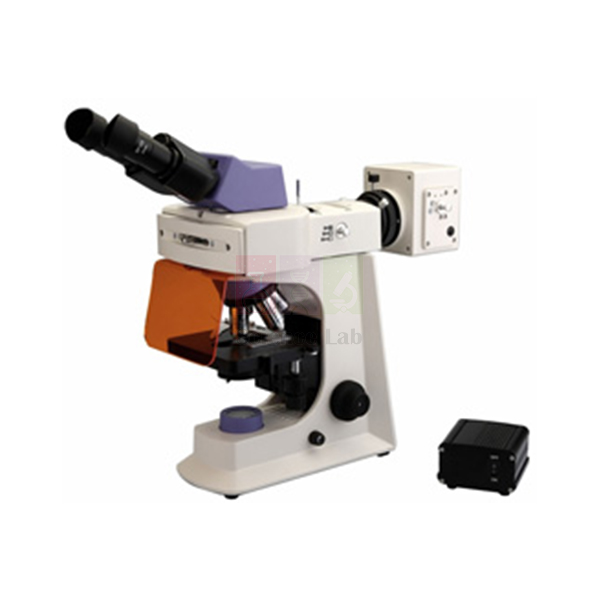
LED Fluorescence Microscope
LED Fluorescence Microscope (Product Code: SLE/M/36) is a trinocular microscope used for both bright-field and fluorescence examination of stained cells, tissues and microorganisms in microbiology, cell biology and research laboratories. It pairs a 4 W LED for transmitted light with 100 W mercury fluorescence illumination and UV, blue and green excitation filters. It is supplied by Jain Scientific Equipments Pvt Ltd (ScienceLab), Ambala, India, for buyers of a fluorescence microscope in India and overseas.
Specifications
| Parameter | Detail |
|---|---|
| Eyepieces | Wide field WF10X; 4 antifungal eyepieces; field of view 20 and 22 mm or higher |
| Eyepiece tube | Trinocular with interpupillary adjustment; 2-way light distribution (100/0 and 0/100) |
| Objectives | Infinity plan achromatic PL 4X/0.10, PL 10X/0.25, PL 40X/0.65 (spring), PL 100X/1.25 (spring, oil) |
| Objective correction | Harmonic-corrected antifungal plan objectives for fluorescence and bright field (10X, 40X, 100X) |
| Nosepiece | Four or more positions, facing inward |
| Stage | Ceramic-coated mechanical X/Y stage, rust free and scratch proof, with vernier calibration; holds two slides |
| Condenser | Upright, universal turret type for bright field and fluorescence imaging; for 10X–100X objectives |
| Transmitted light source | LED 4 W, uniform light distribution, intensity controller, life span of more than 40,000 hours |
| Fluorescence illumination | 100 W mercury, with intensity controller |
| Excitation filters | Three or more (UV, blue and green) in a four-position rotating filter turret |
| Power supply | 12 V DC, 1.5 A, for mains 100–240 V AC, 47–63 Hz, with voltage stabiliser |
| Product code | SLE/M/36 |
| Supplied by | Jain Scientific Equipments Pvt Ltd (ScienceLab), Ambala, India |
What Is the Fluorescence Microscope Used For?
Fluorescence microscopy makes specific structures glow against a dark background: a dye or fluorescent label absorbs light of one colour and emits another, so nuclei, bacteria or labelled proteins stand out clearly. Laboratories use it for fluorescent staining practicals, immunofluorescence teaching and research on cells and microorganisms, then switch the same instrument back to ordinary bright field for routine stained slides.
Choose the UV, blue or green excitation filter on the four-position turret to match the dye, and set the mercury lamp intensity with its controller. The universal turret condenser moves between bright-field and fluorescence settings, and the 2-way light path sends all light to the eyepieces or all to the camera port. Keep the room dim during fluorescence work so faint signals remain visible.
What to Specify When Ordering
- The dyes you use, so the listed UV, blue and green filters can be checked against them
- Whether you need a camera for the trinocular port (not included in the listed specification)
- Your mains voltage and frequency (listed for 100–240 V AC, 47–63 Hz)
- Quantity and any tender specification to be matched
Supplier
ScienceLab, the trading name of Jain Scientific Equipments Private Limited, supplies the LED Fluorescence Microscope from Ambala, Haryana, to universities, research institutes, dealers and importers in India and international markets. For vendor registration, the supplying company's details are CIN U29309HR2020PTC087597 and GSTIN 06AAECJ8618A1ZT. The ScienceLab company profile describes the business.
Related Laboratory Equipment
Consumables and accessories used with a fluorescence microscope:
- Immersion Oil, 25ml — for the 100X/1.25 oil objective; ask us about suitability for fluorescence
- Microscope Slides Glass — for preparing stained specimens
- Cover Glasses — to cover mounted preparations
- Microscope Digital Camera, 5MP — for recording images from the trinocular port; confirm the fit
Browse more instruments in our Microscope category.
Frequently Asked Questions
What light sources does the LED Fluorescence Microscope use?
The LED Fluorescence Microscope uses a 4 W LED with intensity control for transmitted bright-field light, listed with a life span of more than 40,000 hours, and a separate 100 W mercury illuminator with its own intensity controller for fluorescence excitation.
Which excitation filters come with the LED Fluorescence Microscope?
The LED Fluorescence Microscope is listed with three or more excitation filters — UV, blue and green — mounted in a four-position rotating filter turret. Tell us the dyes or labels you plan to use so the filter set can be checked before quotation.
What objectives does the LED Fluorescence Microscope have?
The LED Fluorescence Microscope has infinity plan achromatic objectives PL 4X/0.10, PL 10X/0.25, PL 40X/0.65 (spring) and PL 100X/1.25 (spring, oil), described as harmonic-corrected and antifungal, on an inward-facing nosepiece of four or more positions.
Can images be captured with this microscope?
The LED Fluorescence Microscope has a trinocular tube with 2-way light distribution, sending 100% of light to the eyepieces or 100% to the camera port. A camera is not part of the listed specification, so request one separately if you need image capture.
What power supply does the LED Fluorescence Microscope need?
The LED Fluorescence Microscope runs from a 12 V DC, 1.5 A supply for mains of 100–240 V AC at 47–63 Hz, with a voltage stabiliser. This range covers most laboratory mains supplies, but state your local voltage in the enquiry.
Who supplies the LED Fluorescence Microscope?
The LED Fluorescence Microscope is supplied by Jain Scientific Equipments Pvt Ltd, trading as ScienceLab, Ambala, Haryana, India. The company handles orders from universities, research institutes, dealers and importers in India and international markets.
Request a Quote
To receive a quotation for the LED Fluorescence Microscope, send your dyes, camera requirement and quantity through the contact page, or add it to the enquiry cart with slides and immersion oil.
Last updated: 24 September 2026
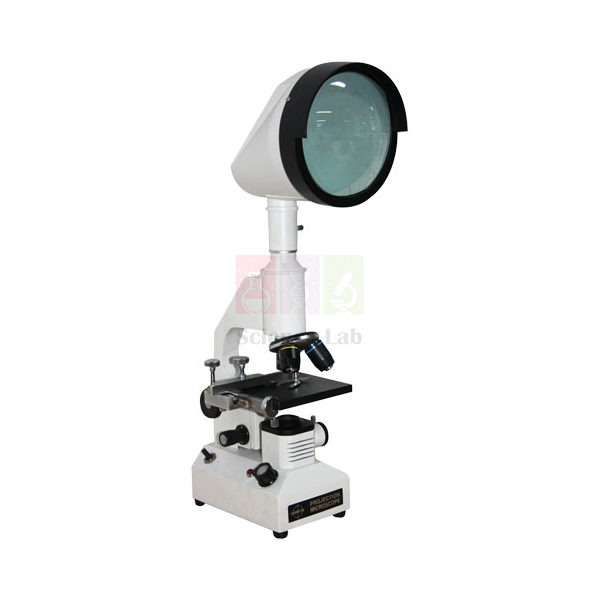
Screen Projection Microscope
Screen Projection Microscope (Product Code: SLE/M/37) is a microscope that projects the magnified image onto a built-in viewing screen, used for group viewing, inspection and angle checks in teaching and laboratory settings. Its 195 mm screen is graduated through 360° and rotates, and objectives from 5x to 60x give 125x to 1000x. It is supplied by Jain Scientific Equipments Pvt Ltd (ScienceLab), Ambala, India, a laboratory equipment supplier in India.
Specifications
| Parameter | Detail |
|---|---|
| Screen diameter | 195 mm |
| Screen | Rotatable, graduated 360° |
| Light control | Solid-state intensity control system |
| Stage | Mechanical stage |
| Objectives | 5x, 10x, 20x, 40x or 60x |
| Magnification | 125x to 1000x |
| Packing | Complete in cardboard box |
| Product code | SLE/M/37 |
| Supplied by | Jain Scientific Equipments Pvt Ltd (ScienceLab), Ambala, India |
What Is the Screen Projection Microscope Used For?
Instead of one person looking into an eyepiece, a projection microscope throws the image onto a screen that several people can see at once. A teacher can point to a cell, fibre or surface defect while a small group watches, and an inspector can compare a part against its expected outline without eye strain during long checking sessions.
Because the screen rotates and carries a 360° scale, angles in the projected image can be read directly: line up a reference with one edge, turn the screen to the next edge and note the change. Adjust brightness with the solid-state intensity control to keep the projected image clear in a normally lit room, and move the specimen with the mechanical stage rather than by hand.
What to Specify When Ordering
- Which objectives you need from the 5x, 10x, 20x, 40x and 60x options
- The magnification range most important for your specimens (125x to 1000x listed)
- Mains voltage and quantity
Supplier
Trading as ScienceLab, Jain Scientific Equipments Private Limited supplies the Screen Projection Microscope from Ambala, Haryana, to schools, colleges, inspection laboratories, dealers and importers in India and international markets. Share your objective choice and we will quote the instrument together with any slides or accessories you need. More about the company is on the profile page.
Related Laboratory Equipment
Accessories and alternatives for projection and measuring work:
- Toolmaker Microscope — for micrometer-based measurement of angles and dimensions
- Microscope Slides Glass — for mounting specimens on the stage
- Lens Tissue — for cleaning the objectives and screen optics
- Carry Case for Microscopes — a sturdier storage option to discuss
Find other models in the Microscope category.
Frequently Asked Questions
How does the Screen Projection Microscope display the image?
The Screen Projection Microscope projects the magnified image onto a built-in screen of 195 mm diameter instead of an eyepiece. Several students or inspectors can view the same specimen together, which makes demonstrations and discussion of what is seen much easier.
What magnification range does the Screen Projection Microscope cover?
The Screen Projection Microscope covers 125x to 1000x on the screen. Objectives are listed as 5x, 10x, 20x, 40x or 60x, so tell us which of these you require and we will quote the matching configuration for your application.
Why is the projection screen rotatable and graduated?
The screen of the Screen Projection Microscope rotates and is graduated through 360°, so it can be used to read angles in the projected image. Align a reference with one edge of the feature, rotate to the second edge and take the difference.
How is the brightness of the image adjusted?
The Screen Projection Microscope has a solid-state intensity control system for its illumination. Raising or lowering the light level keeps the projected image readable at different magnifications and in rooms with different amounts of ambient light.
How is the Screen Projection Microscope packed?
The Screen Projection Microscope is supplied complete in a cardboard box. If you need a sturdier case for frequent moves between rooms or for long-term storage, ask about a carry case when you request your quotation.
Who supplies the Screen Projection Microscope?
The Screen Projection Microscope is supplied by Jain Scientific Equipments Pvt Ltd, trading as ScienceLab, Ambala, Haryana, India, to schools, colleges, inspection laboratories, dealers and importers in India and international markets.
Request a Quote
Tell us the objectives and quantity you need for the Screen Projection Microscope through the contact page, or add it to the enquiry cart, and we will send a quotation.
Last updated: 24 September 2026
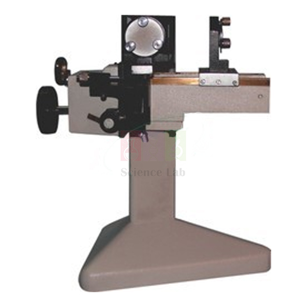
Michelson Interferometer
Michelson Interferometer (Product Code: SLE/M/38) is an optical instrument used to produce and measure interference fringes, for experiments such as finding the wavelength of monochromatic light in college physics laboratories. Its movable mirror is driven by a 1 mm pitch lead screw, with a slow-motion drum reading to 0.0001 mm. It is made by Jain Scientific Equipments Pvt Ltd (ScienceLab), an OEM manufacturer, at its own factory in Ambala, India, for buyers of optics lab equipment in India and abroad.
Specifications
| Parameter | Detail |
|---|---|
| Mirrors | Movable surface-coated mirror; fixed surface-coated mirror loaded with springs |
| Optics | Beam splitter and compensator |
| Mirror no. 1 drive | Main knob, least count 0.01 mm |
| Mirror no. 2 drive | Slow-motion drum, least count 0.0001 mm |
| Lead screw | Well-ground, pitch 1 mm, with spring knob for adjustment |
| Mirror alignment | 3 knurled heads to set mirror no. 2 parallel to mirror no. 1 |
| Pin-hole adjustment | Screws with nut and spring for horizontal and vertical adjustment of the pin-hole view |
| Viewing | Universal adjustable telescope with cross-line graticule; telescope holding clamp (universal type); adjustable eyepiece |
| Illumination optics | Diffused glass screen with pin-hole holder; pin-hole disc; adjustable condenser unit |
| Scale reading | Viewing window to read the main scale |
| Mounting | Base on stand for the interferometer |
| Product code | SLE/M/38 |
| Manufacturer | Jain Scientific Equipments Pvt Ltd (ScienceLab) — OEM |
| Country of origin | India (made in Ambala, Haryana) |
What Is the Michelson Interferometer Used For?
The beam splitter divides light from the source into two paths, one to each mirror; when the beams recombine they form circular fringes seen through the telescope. Moving mirror no. 1 changes one path length, and every fringe that appears or vanishes at the centre corresponds to half a wavelength of travel. Counting fringes against the drum reading gives the wavelength of a monochromatic source, a standard experiment for B.Sc. and M.Sc. physics.
The same set-up is used to find the separation of the sodium D lines from the spacing of fringe visibility, and to estimate the thickness or refractive index of a thin transparent sheet placed in one arm. Start by aligning with the pin-hole disc and the three knurled heads until the two pin-hole images coincide, then switch to the diffused screen and bring the fringes into view before taking readings. It is one of the classic Michelson interferometer India syllabus experiments.
What to Specify When Ordering
- Number of interferometers for the optics laboratory
- Whether you also need a light source, such as a sodium lamp and its power supply (not part of the listed parts)
- Any spare parts you want with the order, such as an extra pin-hole disc
OEM Manufacturer
Jain Scientific Equipments Private Limited, trading as ScienceLab, is the OEM (original equipment manufacturer) of this Michelson Interferometer. The instrument is made and assembled at the company's own licensed factory at 449, HSIIDC Industrial Area, Saha, Ambala, Haryana, India, rather than bought in from a trader.
Because the manufacturer quotes directly, universities, colleges, dealers and importers in India and international markets deal with the people who build the interferometer. Replacement parts such as the pin-hole disc, eyepiece or telescope clamp, and repeat orders for additional lab benches, are handled by the manufacturer itself — just ask us. Read more on the ScienceLab company profile.
Related Laboratory Equipment
Light sources and optics apparatus used with an interferometer:
- Sodium Spectral Lamp — the usual monochromatic source for wavelength and D-line experiments
- Spectral Lamp Power Supply with Lamp Holder — to run the spectral lamp
- Semiconductor Laser — a bright source that can help with first alignment
- Spectrometer — for related wavelength and prism experiments
This instrument is listed in our Microscope category; more optics apparatus is in Physics Lab Equipment.
Frequently Asked Questions
What experiments can be done with the Michelson Interferometer?
The Michelson Interferometer is used to determine the wavelength of monochromatic light, the separation of the sodium D lines, and the thickness or refractive index of a thin transparent sheet. These are standard experiments in undergraduate and postgraduate physics practicals.
How finely can the mirror be moved on the Michelson Interferometer?
On the Michelson Interferometer, the main knob moves mirror no. 1 with a least count of 0.01 mm, and a slow-motion drum gives a least count of 0.0001 mm. Both act through a well-ground lead screw of 1 mm pitch, with the main scale read through a viewing window.
How are the mirrors of the Michelson Interferometer aligned?
Alignment on the Michelson Interferometer uses three knurled heads that set mirror no. 2 parallel to mirror no. 1, together with the pin-hole disc and diffused glass screen. Spring-loaded screws adjust the pin-hole view horizontally and vertically until circular fringes appear.
Is a light source included with the Michelson Interferometer?
A light source is not among the listed parts of the Michelson Interferometer, which includes a condenser unit, pin-hole disc and diffused screen for the illumination path. Most laboratories use a sodium spectral lamp with its power supply; add one to your enquiry if needed.
Who manufactures the Michelson Interferometer?
The Michelson Interferometer is manufactured by Jain Scientific Equipments Pvt Ltd (ScienceLab) as the OEM, at its own licensed factory at Saha, Ambala, Haryana, India. Quotations, repeat orders and replacement-part requests go directly to the manufacturer rather than through a trader.
Is a calibration certificate available for the Michelson Interferometer?
Calibration-certificate availability for the Michelson Interferometer should be discussed with us before ordering. Mention it in your enquiry through the contact page and we will confirm what documentation can be provided with the instrument.
Request a Quote
Quotations for the Michelson Interferometer come straight from the manufacturer. Send the quantity and any light source or spares you need through our contact page, or add the item to the enquiry cart.
Last updated: 24 September 2026
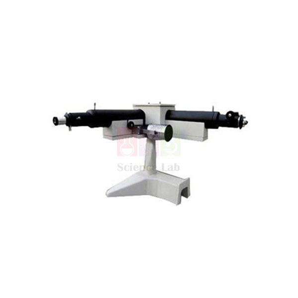
Constant Deviation Spectrograph
Constant Deviation Spectrograph (Product Code: SLE/M/39) is a prism spectroscopic instrument used to observe, measure and record spectral lines in advanced physics laboratories. Its Broca-type prism is turned by an external wavelength drum, so the collimator and telescope stay fixed while each wavelength is brought into view. It is built by Jain Scientific Equipments Pvt Ltd (ScienceLab), an OEM manufacturer, at its own factory in Ambala, India, as part of its physics lab equipment in India.
Specifications
| Parameter | Detail |
|---|---|
| Base | Heavy, stable base with pillar carrying a platform for the collimator, telescope and prism housing |
| Prism table | Broca-type prism, refractive index 1.65/1.74, housed under a cover and rotated by an outside wavelength drum |
| Telescope | Mounted on the platform, rack-and-pinion focusing, achromatic objective, eyepiece with scale (least count 0.01 cm) |
| Collimator | Achromatic objective, rack-and-pinion focusing, slit opening varied by micrometer screw |
| Prisms | Glass prism E.D.F.; glass prism D.E.D.F. |
| Extension mount | For L.G. plate, Edser-Butler plate or Fabry-Perot etalon |
| Telescope attachments (extra) | Monochromator conversion attachment; filar micrometer eyepiece reading 0.001 cm; camera with plate holder; plate holder for camera |
| General accessories | Standard accessory bar, nearly 3 feet long; Fabry-Perot etalon, 30 mm aperture, separation 3, 5 or 10 mm as desired; Edser-Butler plate for determining wavelength; L.G. plate with stand; condenser on stand |
| Product code | SLE/M/39 |
| Manufacturer | Jain Scientific Equipments Pvt Ltd (ScienceLab) — OEM |
| Country of origin | India (made in Ambala, Haryana) |
What Is the Constant Deviation Spectrograph Used For?
In an ordinary spectrometer the telescope swings round to follow each colour. In a constant deviation instrument the collimator and telescope are fixed at one angle and the prism rotates instead; turning the wavelength drum brings successive lines onto the cross-wire so that wavelengths can be read directly. Physics departments looking for a prism spectrometer in India choose this design for M.Sc. work on line spectra of sodium, mercury and other discharge lamps.
The optional parts extend the experiments. A Fabry-Perot etalon or Lummer-Gehrcke (L.G.) plate on the extension mount reveals the fine structure of a line, the Edser-Butler plate measures wavelength from channelled spectra, the filar micrometer eyepiece measures small line separations to 0.001 cm, and the camera with plate holder records a spectrum. With the monochromator attachment the instrument can also deliver light of a selected wavelength.
What to Specify When Ordering
- Prism: glass E.D.F. or D.E.D.F.
- Telescope attachments: monochromator attachment, filar micrometer eyepiece, camera and plate holder
- Fabry-Perot etalon separation: 3 mm, 5 mm or 10 mm
- Other accessories: accessory bar, Edser-Butler plate, L.G. plate with stand, condenser on stand
- Number of instruments
OEM Manufacturer
ScienceLab is the trading name of Jain Scientific Equipments Private Limited, the OEM (original equipment manufacturer) of this Constant Deviation Spectrograph. It is produced at the company's own licensed factory in the HSIIDC Industrial Area at Saha, Ambala, Haryana, India, where the complete instrument is made and assembled rather than bought in from another supplier.
Buying from the maker means the configuration you order — prism, etalon separation and attachments — is quoted and supplied factory-direct to universities, research institutes, dealers and importers in India and international markets. Additional accessories or replacement parts for instruments already in your laboratory can be requested from the manufacturer; ask us. See the ScienceLab profile.
Related Laboratory Equipment
Sources and optics apparatus used in spectroscopy practicals:
- Sodium Spectral Lamp — a common line source for calibration and fine-structure work
- Spectral Lamp Power Supply with Lamp Holder — powers the spectral lamp
- Spectrometer — for introductory prism and grating experiments
- Michelson Interferometer — for interference-based wavelength measurement
The instrument is listed in our Microscope category; further optics apparatus is in Physics Lab Equipment.
Frequently Asked Questions
How does the Constant Deviation Spectrograph work?
The Constant Deviation Spectrograph keeps its collimator and telescope fixed and rotates a Broca-type prism with an outside wavelength drum. Each wavelength in turn is deviated through the same angle into the telescope, so spectral lines are brought to the cross-wire and read from the drum.
Which prisms are used in the Constant Deviation Spectrograph?
The Constant Deviation Spectrograph has a Broca-type prism of refractive index 1.65/1.74 housed under a cover on the prism table. Glass prisms in E.D.F. and D.E.D.F. grades are also listed, so state which you require when you ask for a quotation.
What telescope attachments are available?
The Constant Deviation Spectrograph offers extra telescope attachments: a conversion attachment that turns it into a monochromator, a filar micrometer eyepiece reading 0.001 cm, a camera with plate holder, and a separate plate holder for the camera.
What Fabry-Perot etalon can be supplied with the spectrograph?
The Fabry-Perot etalon offered with the Constant Deviation Spectrograph has a 30 mm aperture and an etalon separation of 3 mm, 5 mm or 10 mm, as desired by the customer. It fits the extension mount, which also takes an L.G. plate or Edser-Butler plate.
Who manufactures the Constant Deviation Spectrograph?
The Constant Deviation Spectrograph is manufactured by Jain Scientific Equipments Pvt Ltd (ScienceLab), the OEM, at its own licensed factory at Saha, Ambala, Haryana, India. Quotations, accessory orders and replacement-part requests are handled directly by the manufacturer.
Is a light source included with the Constant Deviation Spectrograph?
A lamp is not among the listed parts of the Constant Deviation Spectrograph, although a condenser on stand is included in the general accessories. Laboratories usually add a spectral lamp and power supply; include them in your enquiry if you need a complete set-up.
Request a Quote
Your quotation for the Constant Deviation Spectrograph comes direct from the manufacturer. List the prism, etalon separation and attachments you need on our contact page, or add the instrument to the enquiry cart.
Last updated: 24 September 2026
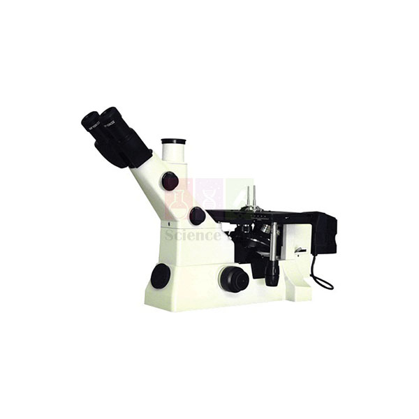
Research Inverted Metallurgical Microscope
Research Inverted Metallurgical Microscope (Product Code: SLE/M/40) is an inverted reflected-light microscope used to examine polished and etched metal specimens in metallurgy, materials science and engineering laboratories. Its objectives look up at the specimen from below a 150 x 200 mm stage, and Koehler illumination from a 6 V 20 W halogen lamp gives even lighting. It is supplied by Jain Scientific Equipments Pvt Ltd (ScienceLab), Ambala, India, a laboratory equipment exporter from India.
Specifications
| Parameter | Detail |
|---|---|
| Observation head | Binocular, with eyepiece tube, inclined at 30° |
| Interpupillary distance | 55 to 75 mm |
| Eyepiece | 10x (16 mm) |
| Objectives | PL10x, PL25x, PL40x; PL60x optional |
| Nosepiece | Quadruple, revolving on ball bearing |
| Mechanical stage | Co-axial, 150 x 200 mm, cross travel 15 x 15 mm |
| Illumination | Koehler illumination system, 6 V 20 W halogen lamp, adjustable brightness |
| Focusing | Co-axial focusing control knobs, tension adjustment ring and pre-focusing lock |
| Main body | All parts die-cast for long life and mechanical strength |
| Product code | SLE/M/40 |
| Supplied by | Jain Scientific Equipments Pvt Ltd (ScienceLab), Ambala, India |
What Is the Inverted Metallurgical Microscope Used For?
Metals are opaque, so their structure is studied with light reflected from a polished and etched surface. Students use this microscope to see grain boundaries, phases such as ferrite and pearlite, inclusions, porosity and weld zones, and to compare heat-treated samples in a materials science practical.
Because the objectives sit under the stage, the prepared face of the specimen simply rests downward on the stage plate. Samples of different heights, including mounted pucks and small cut sections, stay in the same focal plane without levelling in plasticine. Start with the PL10x objective to find the area, then move to PL25x or PL40x for detail, and use the 15 x 15 mm cross travel to scan the surface systematically.
What to Specify When Ordering
- Whether the optional PL60x objective is required
- Any imaging requirement (the listed head is binocular), so we can advise before quoting
- Mains voltage and number of microscopes
Supplier
Jain Scientific Equipments Private Limited supplies this microscope under its ScienceLab trading name from Ambala, Haryana, to engineering colleges, metallurgy departments, industrial training centres, dealers and importers in India and international markets. For supplier onboarding and import paperwork, its registrations are GSTIN 06AAECJ8618A1ZT and Importer-Exporter Code AAECJ8618A issued by DGFT. The ScienceLab profile gives more background.
Related Laboratory Equipment
Specimen preparation and testing equipment used alongside metallography:
- Single Disc Polishing Machine — to prepare the flat, polished specimen face
- Vickers Hardness Tester — hardness readings to relate to the observed microstructure
- Brinell Hardness Tester — bulk hardness testing of the same materials
- Research Trinocular Ore Microscope — for ore and mineral specimens
See the Microscope category for more instruments, and Materials Testing and Properties for testing equipment.
Frequently Asked Questions
Why is the Research Inverted Metallurgical Microscope built upside down?
The Research Inverted Metallurgical Microscope places its objectives beneath the stage, so a polished metal specimen rests face down on the stage plate. Specimens of different heights stay in focus without levelling, and heavy or irregular samples can be examined without special mounting.
What objectives and eyepiece does the Research Inverted Metallurgical Microscope have?
The Research Inverted Metallurgical Microscope has PL10x, PL25x and PL40x objectives, with PL60x available as an option, on a quadruple nosepiece that revolves on a ball bearing. The eyepiece is 10x (16 mm) in a binocular head inclined at 30°.
What illumination does this metallurgical microscope use?
The Research Inverted Metallurgical Microscope uses a Koehler illumination system with a 6 V 20 W halogen lamp and adjustable brightness. Koehler lighting spreads light evenly over the field, which helps when judging grain structure and etching contrast across a polished surface.
How big is the stage and how far does it travel?
The Research Inverted Metallurgical Microscope has a co-axial mechanical stage measuring 150 x 200 mm with a cross travel of 15 x 15 mm. The large stage supports bulky specimens, and the controlled travel lets the user scan the prepared surface field by field.
Can a camera be used with the Research Inverted Metallurgical Microscope?
The Research Inverted Metallurgical Microscope is listed with a binocular observation head, and no camera port is included in its specification. If you need to capture micrographs, mention this in your enquiry so we can advise on suitable options before quoting.
Who supplies the Research Inverted Metallurgical Microscope?
The Research Inverted Metallurgical Microscope is supplied by Jain Scientific Equipments Pvt Ltd, trading as ScienceLab, Ambala, Haryana, India. It serves engineering colleges, metallurgy departments, training centres, dealers and importers in India and international markets.
Request a Quote
Request a quotation for the Research Inverted Metallurgical Microscope by sending your objective choice and quantity through our contact page, or add it to the enquiry cart with polishing and hardness-testing equipment.
Last updated: 24 September 2026
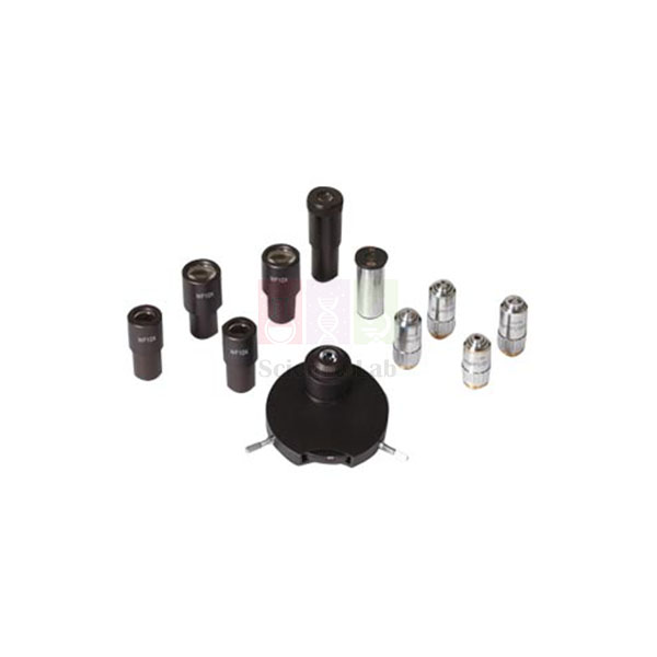
Phase Contrast & Dark Field For Microscopes
Phase Contrast & Dark Field For Microscopes (Product Code: SLE/M/41) is a turret phase-contrast attachment set used to make transparent, unstained specimens visible in biology, microbiology and research laboratories. It comprises four plan phase objectives and a precision turret condenser with matching annuli, plus a zero position for ordinary bright field. It is supplied by Jain Scientific Equipments Pvt Ltd (ScienceLab), Ambala, India, for labs adding microscope phase contrast in India and abroad.
Key Details
| Parameter | Detail |
|---|---|
| Type | Turret phase contrast equipment |
| Objectives | Four plan phase objectives: PLPH-10x, PLPH-20x/25x, PLPH-40x, PLPH-100x |
| Condenser | Precision turret condenser unit with optical annuli matched to all four plan phase objectives |
| Bright-field use | Neutral "zero position" on the turret for normal bright-field microscopy |
| Supplied with | Centering telescope and green filter |
| Dark field | Named in the product title; please confirm the dark-field arrangement with us |
| Product code | SLE/M/41 |
| Supplied by | Jain Scientific Equipments Pvt Ltd (ScienceLab), Ambala, India |
What Is a Phase Contrast Attachment Used For?
Living cells, protozoa, cheek epithelium and many bacteria are almost transparent, so under ordinary bright field they look faint unless they are stained — and staining usually kills them. Phase contrast turns the tiny delays that light suffers passing through different parts of a cell into differences of brightness, so nuclei, membranes and organelles appear with clear contrast in living, unstained material.
To set up, turn the condenser turret to the annulus that matches the phase objective in use, then look through the centering telescope in place of an eyepiece and centre the bright ring on the dark phase ring. Place the green filter in the light path for a crisper image. Turning the turret to the zero position returns the microscope to normal bright-field work for stained slides.
What to Specify When Ordering
- The make and model of the microscope the set will be fitted to, so compatibility can be checked
- Your choice of 20x or 25x for the second phase objective
- Whether you need a dark-field facility, so the arrangement can be confirmed
- Number of sets
Supplier
This phase contrast set is supplied by Jain Scientific Equipments Private Limited, which does business as ScienceLab, from Ambala, Haryana. Colleges, hospitals' teaching laboratories, research institutes, dealers and importers in India and international markets can order it on its own or together with a microscope and consumables. See the ScienceLab company profile for more.
Related Laboratory Equipment
Microscopes and consumables used with a phase contrast set:
- Research Binocular Microscope — a research microscope to discuss pairing with the set
- Universal Research Microscope — another binocular instrument; ask us about fit
- Microscope Slides Glass — for wet mounts of living cells
- Cover Glasses — a thin, even cover helps phase images
More options are in our Microscope category.
Frequently Asked Questions
What is included in the Phase Contrast & Dark Field For Microscopes set?
The Phase Contrast & Dark Field For Microscopes set includes four plan phase objectives (PLPH-10x, PLPH-20x/25x, PLPH-40x and PLPH-100x), a precision turret condenser with matching annuli, a centering telescope and a green filter.
Can the microscope still be used for ordinary bright field?
Yes. The turret condenser in the Phase Contrast & Dark Field For Microscopes set has a neutral zero position. Turning to it removes the phase annulus from the light path, so the same microscope can be used for normal bright-field viewing of stained slides.
What is the centering telescope used for?
The centering telescope supplied with the Phase Contrast & Dark Field For Microscopes set replaces an eyepiece during set-up. It lets you see the condenser annulus and the phase ring of each objective so they can be aligned; good alignment is what produces a clear phase-contrast image.
Which microscopes does this phase contrast set fit?
Compatibility of the Phase Contrast & Dark Field For Microscopes set is not stated in its listing. Send the make and model of your microscope through the contact page so we can confirm the fit before you place an order.
Does the set include dark-field equipment?
The Phase Contrast & Dark Field For Microscopes set names dark field in its title, but its listed specification describes only the phase-contrast turret, objectives, centering telescope and green filter. Please confirm the dark-field arrangement with us when you request a quotation.
Who supplies the Phase Contrast & Dark Field For Microscopes set?
The Phase Contrast & Dark Field For Microscopes set is supplied by Jain Scientific Equipments Pvt Ltd, trading as ScienceLab, Ambala, Haryana, India, to colleges, research institutes, dealers and importers in India and international markets.
Request a Quote
Send your microscope model, objective choice and quantity for the Phase Contrast & Dark Field For Microscopes set through our contact page, or add it to the enquiry cart to receive a quotation.
Last updated: 24 September 2026
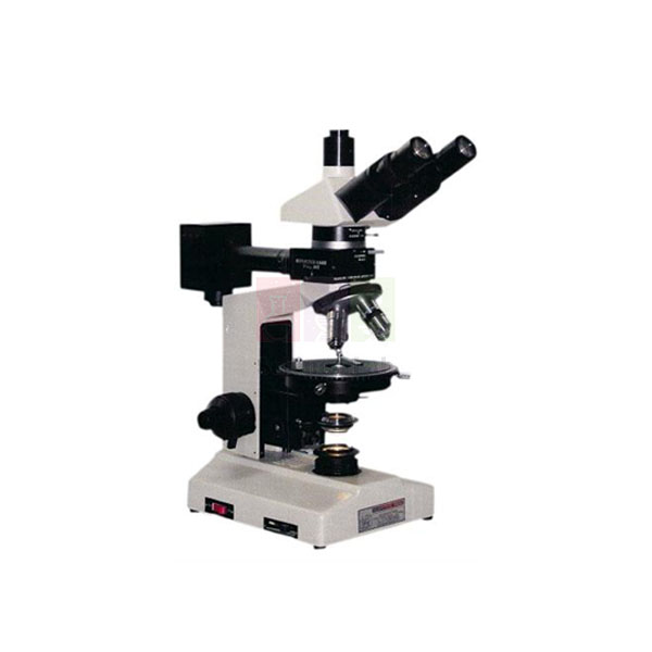
Ore Polarising Microscope
Ore Polarising Microscope (Product Code: SLE/M/42) is a trinocular polarising microscope used to identify ore minerals and study rock specimens in reflected and transmitted polarised light in geology and mining laboratories. It is fitted with two focusable 10X wide-field eyepieces carrying a double cross line, plan POL objectives from 4X to 40X and a colour CCD camera on the trinocular tube. It is supplied by Jain Scientific Equipments Pvt Ltd (ScienceLab), Ambala, India, a laboratory equipment supplier in India.
Specifications
| Parameter | Detail |
|---|---|
| Frame | Transmission and reflection mode polarisation microscopy, built-in filter holders |
| Objectives (plan achromatic, polarising) | 4X POL N.A. 0.13, W.D. 17.0 mm; 10X POL N.A. 0.3, W.D. 10.0 mm; 20X POL N.A. 0.5, W.D. 2.1 mm; 40X POL N.A. 0.75, W.D. 1.1 mm |
| Eyepieces | Wide-field 10X, 2 numbers, with double cross line, focusable |
| Tube | Fixed trinocular tube |
| Light source | High-power LED light with power supply; built-in transmitted light filters |
| Nosepiece | Minimum 4-position, centrable and detachable |
| Focus | Stroke minimum 22 mm; fine stroke minimum 100 µm |
| Condenser | Strain-free polarising condenser, swing-out type |
| Stage | Circular, rotatable and centerable mechanical stage, graduated through 360° |
| Reflected illumination | Polariser slider for the reflected illumination tube |
| Rotating scale | 360° rotatable, scale interval 5° |
| Compensator | Wedge shaped, measurement range 0–4λ |
| Camera | Colour CCD camera, minimum 5 megapixel, on the trinocular tube with a suitable mounting adapter |
| Camera sensor | 2/3 inch CCD; resolution 1920 x 1080 pixels; pixel size 3.45 x 3.45 µm |
| Product code | SLE/M/42 |
| Supplied by | Jain Scientific Equipments Pvt Ltd (ScienceLab), Ambala, India |
What Is the Ore Polarising Microscope Used For?
Ore minerals such as sulphides and oxides are opaque, so a geologist identifies them from how they reflect light: their colour, relative brightness, and whether they change as the stage turns under crossed polars. This instrument provides that reflected polarised view through the polariser slider on the reflected illumination tube, and also a transmitted polarised path for the transparent gangue minerals in a thin section.
The double cross line in each focusable eyepiece is the working reference: centre a grain on the cross, rotate the graduated stage and note where it goes dark to measure extinction positions. The 0–4λ wedge compensator assists with interference-colour work in transmitted light, and the CCD camera records the field for mineral reports or class discussion.
What to Specify When Ordering
- Whether the colour CCD camera is needed or the microscope alone
- Objective set required from the 4X, 10X, 20X and 40X POL objectives
- Mains voltage for the LED power supply
- Quantity, and any tender specification to be met
Supplier
The Ore Polarising Microscope is supplied from Ambala, Haryana, by Jain Scientific Equipments Private Limited under its ScienceLab name. Geology departments, mining institutes, dealers and importers in India and international markets can send their specification and receive a quotation for the configuration they need. The company profile has further details.
Related Laboratory Equipment
Instruments and preparation equipment for ore and mineral microscopy:
- Advance Research Polarising Microscope with Incident Light — infinity-corrected alternative with imaging software
- Research Trinocular Ore Microscope — with a vernier-read round stage and compensator set
- Mineral Test Kit — hand-specimen tests to complement microscope identification
- Single Disc Polishing Machine — for preparing polished sections
Browse the Microscope category for other polarising and research models.
Frequently Asked Questions
What specimens can the Ore Polarising Microscope examine?
The Ore Polarising Microscope works in both transmission and reflection modes. Reflected polarised light is used for opaque ore minerals in polished sections, while transmitted polarised light is used for transparent minerals in thin sections of the same rock.
What eyepieces are supplied with the Ore Polarising Microscope?
The Ore Polarising Microscope comes with two wide-field 10X eyepieces, each with a double cross line and focusing. Focusing the eyepiece makes the cross line sharp for each user, so grains can be centred and angles judged accurately.
What camera is specified for the Ore Polarising Microscope?
The Ore Polarising Microscope is specified with a colour CCD camera of minimum 5 megapixels on the trinocular tube, using a suitable mounting adapter. Its sensor is 2/3 inch, with 1920 x 1080 pixel resolution and 3.45 x 3.45 µm pixels.
Which objectives does the Ore Polarising Microscope use?
The Ore Polarising Microscope uses plan achromatic polarising objectives: 4X POL (N.A. 0.13, W.D. 17.0 mm), 10X POL (N.A. 0.3, W.D. 10.0 mm), 20X POL (N.A. 0.5, W.D. 2.1 mm) and 40X POL (N.A. 0.75, W.D. 1.1 mm).
What focusing and nosepiece features does it have?
The Ore Polarising Microscope has a focus stroke of at least 22 mm with a fine stroke of at least 100 µm, and a centrable, detachable nosepiece of at least four positions, so each objective can be centred to the rotating stage axis.
Who supplies the Ore Polarising Microscope?
The Ore Polarising Microscope is supplied by Jain Scientific Equipments Pvt Ltd, trading as ScienceLab, Ambala, Haryana, India. It is offered to geology departments, mining institutes, dealers and importers in India and international markets.
Request a Quote
For a quotation on the Ore Polarising Microscope, share your camera requirement, objectives and quantity through our contact page, or place it in the enquiry cart.
Last updated: 24 September 2026
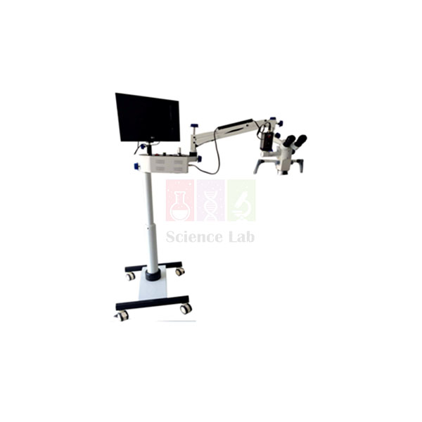
ENT Operating Microscope
ENT Operating Microscope (Product Code: SLE/M/43) is a floor-standing binocular operating microscope used to give a magnified, illuminated view of the ear, nose and throat field in hospital ENT departments and medical teaching settings. Its apochromatic optics have a five-step magnification changer covering 2.4x–40x or more, and an integrated full HD (1080p) camera records video and photos. It is supplied by Jain Scientific Equipments Pvt Ltd (ScienceLab), Ambala, India, a laboratory equipment supplier in India that also serves hospital buyers.
Specifications
| Parameter | Detail |
|---|---|
| Optics | Apochromatic, five-step magnification changer |
| Magnification range | 2.4x–40x or more |
| Maximum field of view | More than 180 mm |
| Objective working distance | f = 250 mm and 400 mm; fine focusing listed for 300 mm |
| Binocular tube | Inclinable, f = 170 mm or higher; interpupillary distance 55–75 mm |
| Eyepieces | Pair of wide-field push-in eyepieces, 10x or 12.5x, with rubber sleeves; diopter setting −5D to +5D |
| Illumination | Coaxial fibre-optic / direct Xenon or LED system; single lamp in LED versions; dual lamp with quick change-over in Xenon versions |
| Light control | Continuous lamp intensity adjustment by a control knob near the surgeon |
| Stand | Stable floor stand on four lockable castors; column height minimum 1.6 m; spring-balance articulated arm, carrier arm, power supply unit |
| Handling | Lateral handgrips for all knobs; dust cover |
| Imaging | Integrated full HD (1080p) camera and full HD recording system with playback for videos and photos |
| Product code | SLE/M/43 |
| Supplied by | Jain Scientific Equipments Pvt Ltd (ScienceLab), Ambala, India |
What Is the ENT Operating Microscope Used For?
ENT work takes place in narrow, deep and poorly lit spaces such as the ear canal and nasal passages. An operating microscope places a bright, coaxial light and a stereoscopic magnified view along the same line of sight, leaving the operator's hands free for instruments. The five-step changer lets the user move from an overview at 2.4x to fine detail at the upper end of the range without refocusing the whole set-up.
The spring-balanced articulated arm holds the head steady in any position, and the lockable castors let staff move the stand between rooms and fix it in place. The integrated full HD camera allows residents and students to follow a procedure on a monitor and lets departments keep recordings for teaching, with playback of stored videos and photos.
What to Specify When Ordering
- Illumination: LED (single lamp) or Xenon (dual lamp with quick change-over)
- Eyepiece magnification: 10x or 12.5x
- Objective working distance required: 250 mm or 400 mm
- Mains voltage and number of units
Supplier
Jain Scientific Equipments Private Limited, trading as ScienceLab, supplies the ENT Operating Microscope from Ambala, Haryana, to hospitals, medical colleges, dealers and importers in India and international markets. Share your specification or tender document and we will quote the matching configuration. Read about the company on the ScienceLab profile page.
Related Laboratory Equipment
Other equipment used in ENT examination rooms and operating theatres:
- Otoscope Set — for routine ear examination before microscope work
- LED Light Otoscope — a handheld examination light for the ear
- Light Operating Room Mobile w/access — general theatre lighting alongside the microscope
- Operating Theatre Table With Accessories — the patient table the microscope stand is positioned beside
This item is listed in our Microscope category; theatre equipment is in Resuscitation & Operating Theatre Equipment.
Frequently Asked Questions
What magnification does the ENT Operating Microscope provide?
The ENT Operating Microscope has apochromatic optics with a five-step magnification changer covering 2.4x–40x or more, and a maximum field of view of more than 180 mm. Eyepieces of 10x or 12.5x are offered, so state your preference when ordering.
What working distances are available on the ENT Operating Microscope?
The ENT Operating Microscope is listed with objective working distances of f = 250 mm and 400 mm, and fine focusing listed for 300 mm. Tell us the working distance your department prefers so the quotation covers the correct objective.
Which illumination options does the ENT Operating Microscope have?
The ENT Operating Microscope uses coaxial fibre-optic or direct illumination with either an LED system, which has a single lamp, or a Xenon system with dual lamps and quick change-over. Intensity is adjusted continuously with a knob placed near the surgeon.
Can the ENT Operating Microscope record video?
Yes. The ENT Operating Microscope includes an integrated full HD (1080p) camera and a full HD recording system. The camera has a playback function for videos and photos, which is useful for teaching, case discussion and departmental records.
What kind of stand does the ENT Operating Microscope use?
The ENT Operating Microscope stands on a stable floor stand with four lockable castors and a column of at least 1.6 m. A spring-balance articulated arm and carrier arm hold the optics, with lateral handgrips on all knobs and a dust cover included.
Who supplies the ENT Operating Microscope?
The ENT Operating Microscope is supplied by Jain Scientific Equipments Pvt Ltd, trading as ScienceLab, Ambala, Haryana, India. It is offered to hospitals, medical colleges, dealers and importers in India and international markets against their specification or tender requirements.
Request a Quote
Send your illumination choice, eyepiece and working distance for the ENT Operating Microscope through our contact page, or add it to the enquiry cart, and we will reply with a quotation.
Last updated: 24 September 2026