- Physics Lab Equipment
- Chemistry Lab Equipment
- Biology Lab Equipment
- Anatomical Lab Equipment
- Learning
- Educational Lab Equipment
- Magnets
- Agriculture
- Astronomy
- Earth Science
- Geography Lab Equipment
- Safety Lab Coat
- Laboratory
- Meteorological
- Electricity & Magnetism
- Heat
- School Lab Equipment
- Lab Glassware
- Science Lab Equipment
- Tvet Lab Equipment
- Robotics Kits Supplier and Manufacturer for Schools India
- Math Lab Equipment Manufacturers and Exporters in Ambala
- STEM Kits Manufacturers and Supplier India
- Engineering Lab Equipment
- Hospital Lab Equipment
- Water, Sanitation & Hygiene Supplies
- Cold Chain, Solar & Communication Equipment
- Nutrition
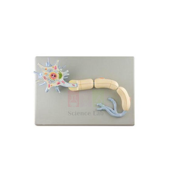
Neuron Model
The Neuron Model (Product Code: SLE/A/147) enlarges a typical myelinated nerve cell about 2,500 times, from the dendrites and cell body to the axon wrapped in its myelin sheath, and shows the organelles inside the cell body. Part of the myelin can be removed to reveal the interior of the axon. Biology teachers, physiology lecturers and psychology departments use it as cell biology equipment in India and elsewhere, and it is made by Jain Scientific Equipments Pvt Ltd (ScienceLab), Ambala.
Specifications
| Parameter | Detail |
|---|---|
| Magnification | About 2,500× life size |
| Size | 53 × 38 × 17 cm |
| Base | Mounted on a durable polymer base |
| Cell body | Nucleus and nucleolus, Golgi complex, mitochondria and endoplasmic reticulum |
| Processes | Dendrites and axon |
| Myelin sheath | With nodes of Ranvier and Schwann cells; a portion can be removed to view the interior of the axon |
| Key | Numbered key included |
Typical Use
The model is ideal for explaining how structure supports function in nerve cells: dendrites receive signals, the cell body with its nucleus, Golgi complex, mitochondria and endoplasmic reticulum keeps the cell supplied, and the axon carries impulses away. Lifting off part of the myelin sheath shows what lies underneath, while the nodes of Ranvier and Schwann cells set up a clear explanation of why myelinated fibres conduct faster.
Teachers often use it straight after an animal cell model lesson, so students can spot familiar organelles in a specialised cell. The numbered key supports labelling worksheets, practical exams and self-study, from senior secondary biology to undergraduate neuroscience.
Manufacturer
Jain Scientific Equipments Private Limited, trading as ScienceLab, builds this neuron model and runs its business from Ambala in Haryana, India. The company profile explains more about who we are.
Related Human Models
Resources for the same lesson on nerve cells and impulses:
- Human Nerve Ending and Synapse Model — follows the axon to the point where the signal passes to the next cell
- Dissectible Human Neuron — a simpler schematic neuron for introductory classes
- Denoyer-Geppert 3-D Human Motor Neuron Model — a motor neuron shown ending on skeletal muscle
- Microslide Nerve & Muscle Action — real microscope images of nerve and muscle to compare with the model
- Micro Slide Viewer — needed to view Microslide image strips
More models are available in our Human Models range.
Frequently Asked Questions
Which cell structures are shown?
The cell body with nucleus and nucleolus, dendrites and axon, Golgi complex, mitochondria, endoplasmic reticulum, and the myelin sheath with nodes of Ranvier and Schwann cells.
Can any part be removed?
Yes. A portion of the myelin sheath can be removed to show the interior of the axon.
Is a key included?
Yes, the model comes with a numbered key.
How is it different from the Dissectible Human Neuron?
This model shows organelle-level detail of a myelinated neuron at about 2,500× on a polymer base, whereas the Dissectible Human Neuron is a schematic nerve cell that separates into parts on a stand.
How large is it?
It measures 53 × 38 × 17 cm, large enough for whole-class demonstration.
Can you supply it for several classrooms or for export?
Yes. We supply schools, universities and distributors in India and international markets; include the quantity with your enquiry.
Request a Quote
Request pricing through our contact page, or add the Neuron Model to the enquiry cart along with any other items you need.
Last updated: 24 September 2026