- Physics Lab Equipment
- Chemistry Lab Equipment
- Biology Lab Equipment
- Anatomical Lab Equipment
- Learning
- Educational Lab Equipment
- Magnets
- Agriculture
- Astronomy
- Earth Science
- Geography Lab Equipment
- Safety Lab Coat
- Laboratory
- Meteorological
- Electricity & Magnetism
- Heat
- School Lab Equipment
- Lab Glassware
- Science Lab Equipment
- Tvet Lab Equipment
- Robotics Kits Supplier and Manufacturer for Schools India
- Math Lab Equipment Manufacturers and Exporters in Ambala
- STEM Kits Manufacturers and Supplier India
- Engineering Lab Equipment
- Hospital Lab Equipment
- Water, Sanitation & Hygiene Supplies
- Cold Chain, Solar & Communication Equipment
- Nutrition
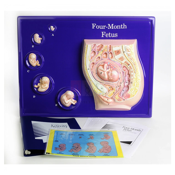
Foetus Model, Four Month
The Foetus Model, Four Month (Product Code: SLE/A/156) presents a 16 week foetus within a model of the womb, with realistic life-sized models of the embryo/foetus at 6, 8, 10, 12 and 14 weeks arranged in sequence beside it. Seeing the stages together at true size helps students grasp how much the developing baby grows during the first four months of pregnancy. Manufactured by Jain Scientific Equipments Pvt Ltd (ScienceLab), Ambala, it is suited to school biology lab equipment collections in India as well as nursing, midwifery and health education classrooms.
Specifications
| Parameter | Detail |
|---|---|
| Main model | 16 week foetus within a model of the womb |
| Earlier stages | Embryo/foetus at 6, 8, 10, 12 and 14 weeks |
| Scale | Realistic, life-sized |
| Arrangement | Earlier stages sequenced next to the 16 week foetus |
| Illustrates | Size, physical features and positioning of the embryo/foetus |
Typical Use
Teachers use the sequence to show the transition from embryo to foetus: learners compare the 6 week stage with the 14 and 16 week stages and describe what changes in body proportions, limbs and facial features. The womb model adds a second lesson on positioning, allowing a discussion of how the foetus lies inside the uterus and how the available space changes as development continues.
Since every stage is life-sized, the model is also effective in health education and antenatal awareness sessions, where real dimensions carry more meaning than enlarged diagrams. Keep it on a stable bench at eye level and let small groups examine it in turn; it then works as hands-on biology lab equipment for reproduction topics rather than a cupboard display.
Manufacturer
This model comes from Jain Scientific Equipments Private Limited, the Ambala, Haryana company known by its trading name ScienceLab, which manufactures teaching models and laboratory equipment for educational institutions. Our company profile gives further background.
Related Human Models
Useful companions for a complete lesson on pregnancy and development:
- Period of Gestation Model — a set of stand-mounted stages that extends the developmental timeline
- Human Embryo Development Set — additional models of early development
- Human Placenta Model — explains how the foetus is nourished during pregnancy
- Human Foetus Model — focuses on the foetus, its membrane and the uterus
- Human Birth Demonstration Model — carries the lesson through to birth
Browse our full Human Models collection for more reproduction and anatomy models.
Frequently Asked Questions
What does “Four Month” refer to?
The main foetus is at 16 weeks, roughly four months of pregnancy, and is shown within a model of the womb. Earlier stages at 6, 8, 10, 12 and 14 weeks are placed alongside it.
Are the models life-sized?
Yes. The embryo and foetus models are described as realistic and life-sized, so students see the real size at each stage.
Does it show how the foetus is positioned?
Yes. Placing the 16 week foetus within the womb model illustrates its positioning as well as its size and physical features.
Can the individual stages be lifted out?
This is not stated in our product documentation. Please contact us if you need the stages to be removable.
Who typically uses this model?
Biology teachers covering human reproduction, and instructors in nursing, midwifery and health education courses who need an accurate sense of early foetal size.
How long does delivery take?
Lead time depends on quantity and destination; share your requirement with us and we will confirm it in the quotation.
Do you export this model?
Yes. We supply institutions and distributors in India and international markets.
Request a Quote
Ask for a quotation on the Foetus Model, Four Month through our contact form, or add it to the enquiry cart with other reproduction models for a combined price.
Last updated: 24 September 2026
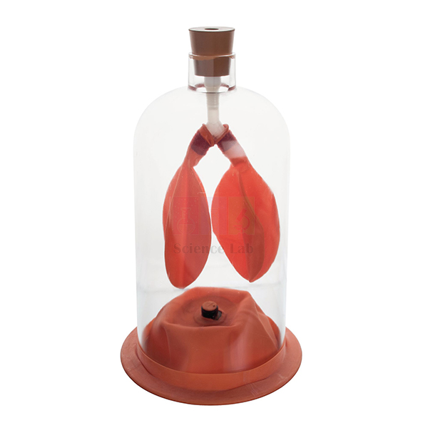
Lung Demonstration Model
The Lung Demonstration Model (Product Code: SLE/A/157) gives students a first-hand view of how the lungs work. Two sacs stand in for the lungs, a rubber membrane acts as the diaphragm and a clear plastic enclosure represents the chest cavity; when the diaphragm is made to relax and contract, the sacs visibly empty and fill with air. Manufactured by Jain Scientific Equipments Pvt Ltd (ScienceLab), Ambala, this simple working model is practical biology lab equipment for respiration lessons in schools across India.
Specifications
| Parameter | Detail |
|---|---|
| Lungs | Two sacs representing the lungs |
| Diaphragm | Rubber membrane |
| Chest cavity | Clear plastic enclosure |
| Size | Height 30 cm x Width 14.5 cm |
| Operation | Diaphragm is made to relax and contract so the lungs empty and fill with air |
Typical Use
When the rubber diaphragm is drawn away from the enclosure, the space around the sacs increases, the pressure inside falls and air flows into the “lungs”, just as it does when the real diaphragm contracts and flattens. Letting the membrane return reverses the change and the sacs deflate. The demonstration links the mechanism of inhalation and exhalation to the pressure–volume relationship taught in physics, so it suits integrated science lessons as well as biology.
Before the lesson, check that the membrane sits evenly and that the sacs inflate freely, then move the diaphragm slowly so the class can match each movement to the change in the sacs. Students can finish by placing a hand on their own abdomen while breathing deeply to feel the same action. Its compact size makes it a sensible early purchase for any biology lab setup in a school.
Manufacturer
The Lung Demonstration Model is made by Jain Scientific Equipments Private Limited, which operates under the ScienceLab name from Ambala, Haryana, and produces teaching equipment for biology, physics and chemistry laboratories. Tender and institutional buyers who need vendor registration details can use GSTIN 06AAECJ8618A1ZT and LEI 3358004HRD1JIVXKYX49. Visit the company profile for more about the company.
Related Laboratory Equipment
Equipment that extends a breathing lesson from mechanism to measurement and anatomy:
- Human Lung Model in Bell Jar — an alternative breathing demonstration in bell-jar form
- Human Respiratory System Model, 2-parts — shows the real anatomy that the demonstration represents
- Lung Volume Bags — lets students collect and compare their own breath volumes afterwards
- Lung Capacity Gauge — for follow-up lung capacity activities
- Segmented Lung — detailed lung anatomy for senior classes
More anatomy teaching models are listed in our Human Models category.
Frequently Asked Questions
How does the model show breathing?
Moving the rubber diaphragm changes the space inside the clear enclosure, so the two lung sacs fill with air and then empty, mirroring inhalation and exhalation.
How big is it?
It stands 30 cm high and is 14.5 cm wide, small enough to demonstrate at the front of a class or pass around a bench.
Is it an anatomically detailed lung model?
No. It is a working demonstration of the breathing mechanism; for detailed structure, use it with an anatomical model such as the Human Respiratory System Model, 2-parts.
Is anything else needed to run the demonstration?
The sacs, rubber diaphragm and enclosure together make up the apparatus, so the basic demonstration can be run with the model alone.
Can a worn rubber membrane be replaced?
Spare-part availability is not listed in our documentation; please contact us to ask about replacement membranes.
Can you supply class sets or export orders?
Yes. We supply schools, colleges and distributors in India and international markets; tell us the quantity and destination for a quotation.
Request a Quote
For prices and availability of the Lung Demonstration Model, get in touch with us or add it to your enquiry cart along with any breathing and respiration equipment you need.
Last updated: 24 September 2026
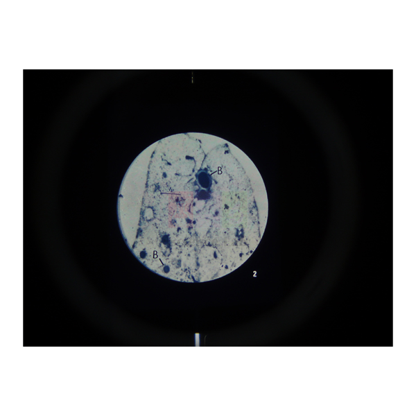
Microslide The Circulatory System
Microslide The Circulatory System (Product Code: SLE/PS/99) is a set of 8 related 35 mm photographs taken through a microscope, used to teach the blood, blood vessels, lymph vessels and heart in secondary biology. Alongside human blood, a phagocyte and an artery with its vein, it includes two views of a rat heart at 4x and 6x and a close-up of the valve at the aorta. It is supplied by Jain Scientific Equipments Pvt Ltd (ScienceLab), Ambala, India.
Specifications
| Parameter | Detail |
|---|---|
| Images per set | 8 related 35 mm images photographed through a microscope |
| Blood | Human Blood; Phagocyte |
| Vessels | Artery and vein; Lung capillaries; Lymph vessel and valves |
| Heart | Rat Heart (4x); Rat heart (6x); Valve at aorta |
| Callouts | Arrows and callouts guide students to key features |
| Folder | Detailed text folder to inform and question students; handy pocket stores the Microslides |
| For use in | Microslide Viewer |
| Product code | SLE/PS/99 |
| Supplied by | Jain Scientific Equipments Pvt Ltd (ScienceLab), Ambala, India |
What Is the Microslide The Circulatory System Used For?
The set gives a class a microscope-level view of the transport system without cutting or staining tissue. A typical sequence starts with human blood and a phagocyte, then compares the thick wall of an artery with its matching vein, and moves to the thin capillaries of the lung where exchange with the air takes place.
The lymph vessel with its valves shows the second, one-way return route for tissue fluid, and the rat heart images at 4x and 6x, followed by the valve at the aorta, help students see why valves stop blood flowing backwards. Viewed in a Microslide Viewer, with callouts on each frame, the set is a practical way to back up a heart model or a glass slide practical.
What to Specify When Ordering
- The number of Microslide The Circulatory System sets
- Whether you also need Microslide Viewers
- Whether a glass slide set of circulatory tissue should be quoted for microscope practicals
Supplier
Microslide The Circulatory System reaches schools, colleges and dealers through Jain Scientific Equipments Private Limited, which operates under the ScienceLab name in Ambala, Haryana. We supply it within a wider range of biology teaching resources for buyers in India and international markets. Visit our company profile for more about who we are.
Related Prepared Slides
These pair well with the set in circulation lessons:
- Microslide Viewer — the viewer the 35 mm images are designed for
- Prepared Microscope Slide Set – Circulatory System — glass slides of artery, vein, cardiac muscle and blood for microscope work
- Human Circulatory System Model — places the vessels in the whole body
- Human Lymphatic System Model — expands on the lymph vessel image
All topic sets are grouped in Prepared Slides.
Frequently Asked Questions
Which images are included in Microslide The Circulatory System?
Microslide The Circulatory System includes human blood, a phagocyte, artery and vein, lung capillaries, a lymph vessel with valves, a rat heart at 4x, a rat heart at 6x, and the valve at the aorta.
How is the heart shown in Microslide The Circulatory System?
Microslide The Circulatory System shows the heart in two rat heart images, at 4x and 6x, so students can study heart structure at low magnification, and adds a separate frame of the valve at the aorta for a closer look at a heart valve.
Does Microslide The Circulatory System replace glass microscope slides?
Microslide The Circulatory System complements rather than replaces glass slides. Its 35 mm images are viewed in a Microslide Viewer with callouts, while glass slides are for students practising their own microscope work.
What student notes come with Microslide The Circulatory System?
Microslide The Circulatory System includes a detailed text folder designed to stimulate, inform and question students about circulation, with a pocket in the folder for storing the Microslides.
Who supplies Microslide The Circulatory System?
Microslide The Circulatory System is supplied by Jain Scientific Equipments Pvt Ltd (ScienceLab), Ambala, Haryana, India, to schools, colleges, dealers and importers, and can be quoted together with models and slide sets.
Request a Quote
Request a quotation for Microslide The Circulatory System on our contact page, giving the quantity and delivery location, or use the enquiry cart to list it with the other items for your lab.
Last updated: 24 September 2026
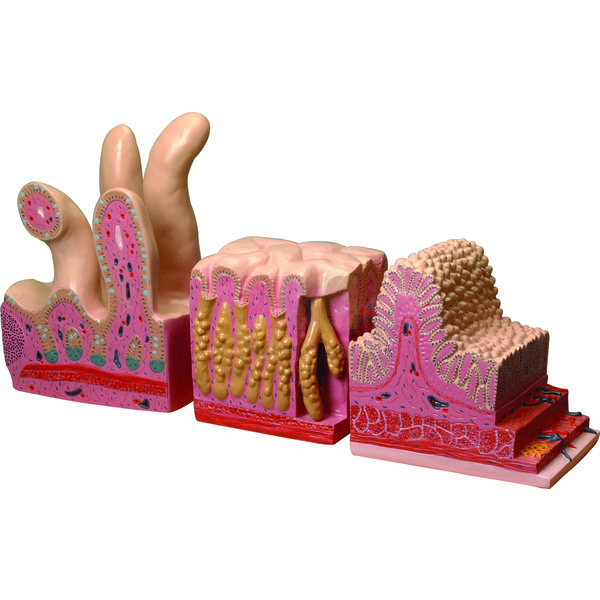
Digestive Canal Model
The Digestive Canal Model (Product Code: SLE/A/158) shows the structure of the digestive canal, the continuous tube through which food passes as it is broken down and absorbed. It is supplied in three parts, and teachers use it to take students through the canal region by region in nutrition and digestion lessons. Made by Jain Scientific Equipments Pvt Ltd (ScienceLab), Ambala, it can form the digestion component of a human body model kit for schools, colleges and nursing institutes in India.
Key Details
| Parameter | Detail |
|---|---|
| Type | Anatomical model of the human digestive canal |
| Shows | Structure of the digestive canal |
| Supplied as | Three parts |
| Suitable for | Digestion and nutrition lessons in schools, colleges and nursing institutes |
Typical Use
Start with the canal as a whole so students appreciate its length and continuity, then focus on individual regions to discuss what each contributes: mechanical and chemical breakdown of food, absorption of nutrients and the recovery of water before waste leaves the body. Asking learners to trace a meal’s journey along the model and name each region in order is a quick check of understanding at the end of the topic.
The model also anchors practical work. After studying it, students can test food samples for starch or protein, or run a starch-digestion test with an enzyme experiment kit, and then point to the region of the canal where that digestion takes place. Linking results to the model helps them remember where each process happens.
Manufacturer
ScienceLab is how Jain Scientific Equipments Private Limited trades; the company is located in Ambala, Haryana, and makes human anatomy models alongside a wider range of school and college laboratory equipment. You can learn more on our company profile page.
Related Laboratory Equipment
Models and slides that support the same digestion lessons:
- Human Digestive System Model — a broader digestive system model for comparison
- Human Digestive Tract Model — an alternative tract model for side-by-side teaching
- Human Stomach Model — closer study of the stomach
- Appendix and Caecum Model — detail of the region where the small and large intestines meet
- Prepared Microscope Slide Set – Digestion — a tissue-level view to accompany the model
Our Human Models range lists further anatomy models for biology departments.
Frequently Asked Questions
What does the model show?
It shows the structure of the human digestive canal, the tube that runs through the body from where food enters to where waste leaves.
How many parts is it supplied in?
Three parts. The arrangement of the parts is shown in the product photograph; contact us for a detailed breakdown.
Does it include the liver and pancreas?
The product documentation describes the digestive canal only. If you need the accessory organs as well, compare the Human Digestive System Model or ask us for advice.
Is a key or labelling supplied?
A key is not mentioned in our documentation, so please confirm with us before ordering if you need one.
Which classes can use it?
It fits the digestion and nutrition topics of middle and secondary school biology and is equally useful for introductory anatomy in nursing and paramedical courses.
Can a school buy several at once?
Yes. Send the quantity and delivery address and we will quote for supply within India or to international markets.
Request a Quote
Request a quotation for the Digestive Canal Model through our contact page, or collect it with other items in the enquiry cart and send one combined enquiry.
Last updated: 24 September 2026
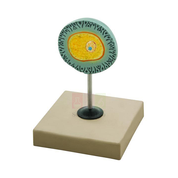
Human Egg Model
The Human Egg Model (Product Code: SLE/A/159) is a teaching model of the human egg, or ovum — the female gamete that joins with a sperm at fertilisation. Mounted on a base so it stands securely on a bench or display shelf, it gives teachers a clear visual starting point for lessons on human reproduction. It is manufactured by Jain Scientific Equipments Pvt Ltd (ScienceLab), Ambala, and is a straightforward item to include when planning a biology lab setup for a school in India.
Key Details
| Parameter | Detail |
|---|---|
| Type | Teaching model of the human egg (ovum) |
| Mounting | Mounted on base |
| Purpose | Visual aid for lessons on the female gamete, ovulation and fertilisation |
| Suitable for | School biology, college zoology, and nursing or health education classes |
Typical Use
A real human egg is barely visible to the naked eye, so a model is the easiest way to discuss it with a group. Teachers use it to explain that the egg is far larger than a sperm cell, that it carries stored nutrients and half of the genetic material a new individual needs, and that it is surrounded by protective layers a sperm must pass through before fertilisation.
Placed next to a sperm model, it lets students contrast the two gametes in size, shape and role; it is also a helpful prop when explaining the menstrual cycle, ovulation and the start of pregnancy in health education sessions. Within a human biology model set it pairs naturally with sperm, embryo and pelvis models.
Manufacturer
Jain Scientific Equipments Private Limited, which trades as ScienceLab, manufactures anatomical teaching models such as this one in Ambala, Haryana, for schools, colleges and distributors. Further information is available on the company profile.
Related Human Models
Models that complete a lesson on gametes and fertilisation:
- Human Sperm Model — the matching male gamete model for comparison lessons
- Embryo Model — shows what develops after fertilisation
- Human Female Genital Organs Model — the organs in which eggs mature and are released
- Female Pelvis Model — a median-section view of the female pelvic organs
Find more reproduction teaching models in our Human Models range.
Frequently Asked Questions
How is the model presented?
It is mounted on a base, so it can stand on a bench, shelf or teacher’s desk during lessons.
Is the egg shown enlarged, and by how much?
Because the human egg is so small, any teaching model of it is enlarged. The exact magnification is not stated in our documentation; contact us if you need it.
Does it come with a key or labelled structures?
This is not listed in our product documentation. Please ask us for details before ordering if labelling is essential.
Can it be quoted together with the Human Sperm Model?
Yes. They are separate items, but you can add both to the enquiry cart and we will quote them together.
How should students handle it?
Lift and carry it by the base rather than the model itself, and keep it away from heat and direct sunlight when stored.
Do you supply schools outside India?
Yes. Orders are shipped to schools, colleges and distributors in India and international markets.
Request a Quote
To get a price for the Human Egg Model, send us an enquiry or add it to the enquiry cart with any companion models.
Last updated: 24 September 2026
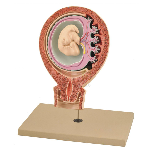
Human Foetus Model
The Human Foetus Model (Product Code: SLE/A/160) is a three-part model that shows the relationship between the foetus, the foetus membrane and the uterus. Rather than tracing growth over time, it concentrates on how the developing baby is enclosed and supported inside the mother, which makes it valuable for reproduction, pregnancy and nursing lessons. It is produced by Jain Scientific Equipments Pvt Ltd (ScienceLab), an anatomical models manufacturer in Ambala, India.
Specifications
| Parameter | Detail |
|---|---|
| Construction | Three-part model |
| Structures shown | Foetus, foetus membrane and uterus |
| Teaching focus | Relationship between the foetus, its membrane and the uterus |
Typical Use
Teachers use the model to explain, from the outside in, how the muscular uterus holds the pregnancy, how the membrane encloses the foetus and cushions it within a fluid-filled space, and how the foetus lies inside this protected environment. Students can then discuss why the membrane matters, what happens when it ruptures at the start of labour, and how the arrangement changes as the pregnancy advances.
In nursing, midwifery and health education courses, the model helps trainees picture what they will later palpate or see on an ultrasound image, and it works well with other biology teaching aids for pregnancy topics, such as placenta and developmental-stage models.
Manufacturer
ScienceLab is the trading name of Jain Scientific Equipments Private Limited (CIN U29309HR2020PTC087597), an Ambala, Haryana company that manufactures at its licensed factory at 449, HSIIDC Industrial Area, Saha, Ambala (Factory Licence AMB-ONLINE-CHD-J-414). See the company profile for details of the business.
Related Human Models
Models that build a fuller picture of pregnancy alongside this one:
- Human Placenta Model — explains the exchange between mother and foetus
- Foetus Model, Four Month — life-sized developmental stages shown within a womb model
- Period of Gestation Model — stand-mounted stages of embryonic and foetal development
- Human Birth Demonstration Model — continues the lesson to the process of birth
View the rest of our Human Models for anatomy and reproduction teaching.
Frequently Asked Questions
What does this model teach?
It shows how the foetus, the foetus membrane and the uterus relate to one another during pregnancy.
How many parts does it have?
It is a three-part model.
How is it different from the Foetus Model, Four Month?
This model focuses on the foetus within its membrane and the uterus, while the Foetus Model, Four Month presents a sequence of life-sized developmental stages beside a foetus in a womb model.
Which stage of pregnancy does the foetus represent, and is it life-size?
Neither is stated in our product documentation; please contact us for these details.
Is a key card included?
A key card is not mentioned in our documentation, so confirm with us if you need one for labelling.
What should a college include in its purchase enquiry?
The quantity, delivery location, billing details and any registration documents your procurement office needs; our company registration details are given above.
Is export supply possible?
Yes, we supply buyers in India and international markets.
Request a Quote
For a quotation on the Human Foetus Model, contact us or add it to your enquiry cart together with other pregnancy and development models.
Last updated: 24 September 2026
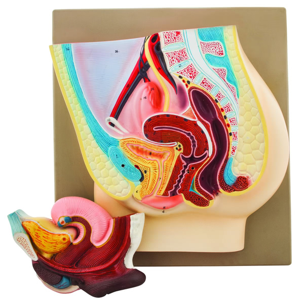
Female Pelvis Model
The Female Pelvis Model (Product Code: SLE/A/161) is a median section of the female pelvis. One half presents the genital organs together with the bladder and a removable rectum, while the other half is shown in its normal position in the pelvis, so students can compare a sectional view with the natural arrangement. A numbered key card is supplied. The model is manufactured by Jain Scientific Equipments Pvt Ltd (ScienceLab), Ambala, one of the human anatomical models manufacturers in India serving schools, colleges and medical training institutes.
Specifications
| Parameter | Detail |
|---|---|
| Section | Median section of a female pelvis |
| First half | Genital organs with bladder and removable rectum |
| Second half | Shown in the normal position in the pelvis |
| Removable part | Rectum |
| Identification | Numbered key card |
Typical Use
In reproductive anatomy classes, the median section lets students see how the internal genital organs sit between the bladder in front and the rectum behind — a relationship that is hard to grasp from surface drawings. Taking out the removable rectum gives a clearer view of the neighbouring structures, and comparing the sectioned half with the half in its normal position helps learners connect a cut-away view with the body as it really is.
Nursing, midwifery and paramedical students use the model to relate pelvic anatomy to clinical topics such as catheterisation, pregnancy and childbirth. The numbered key card makes it easy to run quick identification tests, which is why it is useful school biology lab equipment for senior classes as well as professional training.
Manufacturer
Jain Scientific Equipments Private Limited manufactures the Female Pelvis Model and sells it under the ScienceLab brand. The company is based in Ambala, Haryana, and supplies anatomy models and laboratory equipment to educational and training institutions. The company profile has more information.
Related Human Models
Models that support the same pelvic and reproductive anatomy lessons:
- Female Half Pelvis Model — an alternative half-pelvis presentation for comparison
- Human Adult Female Pelvis Structural Model — the bony pelvis for skeletal context
- Human Female Urogenital Model — brings the urinary and reproductive organs together
- Male Pelvis Section Model — the male counterpart for comparison lessons
See all models in our Human Models category.
Frequently Asked Questions
What does the median section show?
One half shows the genital organs with the bladder and a removable rectum; the other half is shown in its normal position in the pelvis.
Which part can be removed?
The rectum is removable.
Is a key supplied?
Yes, the model comes with a numbered key card for identifying the structures.
Is it natural size?
The scale is not stated in our product documentation; please contact us if you need dimensions before ordering.
Is it suitable for nursing and midwifery training?
Yes. The sectional view of the pelvic organs and the key card suit anatomy teaching in nursing, midwifery and paramedical courses as well as senior school biology.
How should it be cleaned and stored?
Dust it with a soft brush or wipe it with a lightly damp cloth, keep the removable rectum with the model, and store it covered away from heat.
Can institutions order in bulk?
Yes. We quote for single units and larger institutional orders in India and international markets.
Request a Quote
To receive a quotation for the Female Pelvis Model, write to us via the contact page or add it to the enquiry cart.
Last updated: 24 September 2026
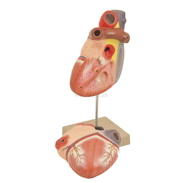
Heart 3 times Natural Size Model
The Heart 3 times Natural Size Model (Product Code: SLE/A/162) is a human heart enlarged 3 times its actual size and dissectible into 2 parts, showing the structure of the auricles and ventricles and, clearly, the chief blood vessels. Made up on a stand 30 cm high, it is large enough to demonstrate at the front of a class yet compact on the bench. It is manufactured by Jain Scientific Equipments Pvt Ltd (ScienceLab), Ambala, as a heart model for anatomy teaching in India’s schools, colleges and medical training institutes.
Specifications
| Parameter | Detail |
|---|---|
| Scale | Enlarged 3 times actual size |
| Construction | Dissectible into 2 parts |
| Internal structure | Auricles and ventricles shown |
| Vessels | Chief blood vessels shown clearly |
| Mounting | Made up on stand |
| Dimensions | Height 30 cm, width 12 cm |
Typical Use
With the model closed, students identify the major vessels entering and leaving the heart; opening it into its two parts reveals the auricles (atria) and ventricles, so the teacher can trace the double circulation — blood returning from the body to the right side, travelling to the lungs, coming back to the left side and leaving for the rest of the body. The threefold enlargement makes the chambers far easier to point out than on a natural-size specimen.
A good routine is to have students label a blank heart diagram first and then check their answers against the model, or to follow a drop of blood through the chambers and vessels aloud. Mounted on its stand, it serves both as demonstration apparatus and as a revision station among school biology lab equipment.
Manufacturer
Made by Jain Scientific Equipments Private Limited, which trades as ScienceLab from Ambala in Haryana, this heart model belongs to a wider range of anatomy models and laboratory apparatus the company produces for educational buyers. Company details are on our company profile.
Related Laboratory Equipment
Heart and circulation items that pair well with this model:
- Human Heart Model — an alternative heart model for comparison
- Human Heart, Pumping Model — demonstrates the pumping action of the heart
- Human Circulatory System Model — places the heart within the whole circulation
- Human Heart on Diaphragm Model — shows where the heart sits in relation to the diaphragm
- Prepared Microscope Slide Set – Circulatory System — slides for microscope work on heart and vessel tissue
Browse the Human Models range for more anatomy models.
Frequently Asked Questions
How much larger than a real heart is it?
It is enlarged 3 times natural size.
How many parts does it come apart into?
It is dissectible into 2 parts, which opens up the auricles and ventricles.
What are the dimensions on the stand?
It stands 30 cm high and is 12 cm wide.
Does it show the heart valves?
Our documentation lists the auricles, ventricles and chief blood vessels. For valve detail, please contact us or refer to the product photograph.
Is a key or label chart included?
A key is not mentioned in the product documentation; ask us if you need one.
Is there a warranty?
Warranty terms are not listed in our documentation and are confirmed with each quotation.
Can you supply several for a department or export them?
Yes. We supply single pieces and multiple units to buyers in India and international markets.
Request a Quote
For pricing on the Heart 3 times Natural Size Model, reach our team or add it to the enquiry cart with other circulation models.
Last updated: 24 September 2026