- Physics Lab Equipment
- Chemistry Lab Equipment
- Biology Lab Equipment
- Anatomical Lab Equipment
- Learning
- Educational Lab Equipment
- Magnets
- Agriculture
- Astronomy
- Earth Science
- Geography Lab Equipment
- Safety Lab Coat
- Laboratory
- Meteorological
- Electricity & Magnetism
- Heat
- School Lab Equipment
- Lab Glassware
- Science Lab Equipment
- Tvet Lab Equipment
- Robotics Kits Supplier and Manufacturer for Schools India
- Math Lab Equipment Manufacturers and Exporters in Ambala
- STEM Kits Manufacturers and Supplier India
- Engineering Lab Equipment
- Hospital Lab Equipment
- Water, Sanitation & Hygiene Supplies
- Cold Chain, Solar & Communication Equipment
- Nutrition
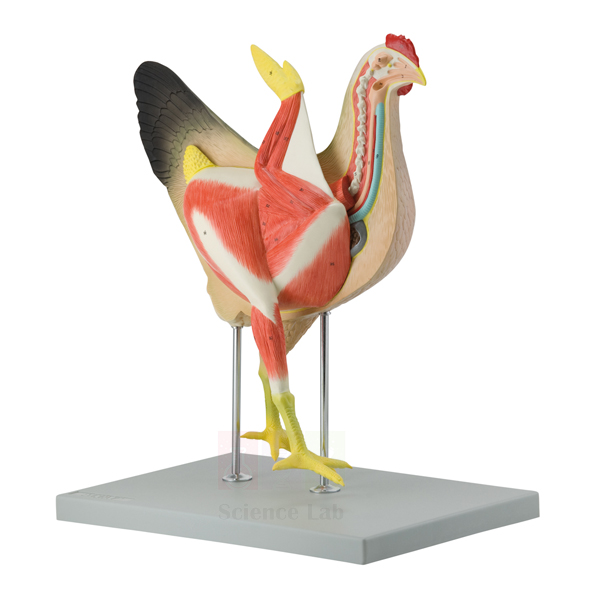
Hen Model
This life-size hen model comes apart into 8 parts on a base. Its right side shows the bird's outer form. The left side is cut lengthwise to expose the muscles, heart, lungs, liver, stomach, intestines and the reproductive system with an egg in place. A numbered key names each structure.
Hen anatomy model specifications
| Parameter | Detail |
|---|---|
| Scale | Life size |
| Construction | 8 parts |
| Size with base | 39.5 x 26 x 46.7 cm |
| Right side | External form of the hen |
| Left side | Longitudinal section showing muscles, heart, liver, intestines, stomach, lungs, reproductive system and an egg |
| Mounting | On a base |
| Supplied with | Numbered key |
| Product code | SLE/A/115 |
| Minimum order | 10 units |
| Dispatch | Usually within 7 to 10 working days |
| Manufacturer | ScienceLab (Jain Scientific Equipments Pvt Ltd), Ambala, India |
Practical work with the hen anatomy model
Outside and inside. Pairs first name the external features on the right side, such as the comb, wattles, beak, wings and shanks. They then turn the model round and list which organs lie under the breast, the back and the vent region, checking each number on the key.
Comparing digestion across classes. Set the hen beside the Mi-Own Frog Model and the Human Digestive System Model. Students trace the gut in each one, from stomach to intestines. They then discuss what a bird uses in place of teeth: a crop that stores food and a muscular gizzard that grinds it.
Flight and circulation. Place a Pigeon Skeleton next to the sectioned side. Students relate the large breast muscles to the keel of the breastbone, then compare the hen's heart with the hearts of other vertebrate groups in the Vertebrate Hearts Model Set.
Egg formation for poultry courses. Use the reproductive system and the egg on the section to describe how a yolk released by the ovary gains albumen, shell membranes and shell on its way down the oviduct. Students then sketch the tract and label each stage.
Frequently Asked Questions About the Hen Model
A hen has no teeth, so how does it break down grain?
Grain is first held and softened in the crop. It then passes to a two-part stomach: the glandular proventriculus adds digestive juices and the muscular gizzard grinds the food, often with grit the bird has swallowed. Point to the stomach on the section while explaining this.
Which tracts open into the cloaca of a hen?
The gut, the ureters from the kidneys and the oviduct all end in one chamber, the cloaca, which opens to the outside at the vent. Droppings, urine wastes and eggs therefore leave by the same exit.
How does a hen's anatomy differ from a mammal's?
Birds have no diaphragm, and their lungs connect to thin air sacs that run through the body and into some bones. A hen has no urinary bladder and passes nitrogen waste as uric acid. Usually only the left ovary and oviduct work.
Which external features should students find on the right side?
Ask them to name the comb, wattles, beak, eye, ear lobe, wing, shank and toes, and to describe how the feathers cover the body. These are the points poultry handlers use when judging the condition of a live bird.
How is the Hen Model taken apart and stored?
It separates into 8 parts so that organs on the sectioned side can be lifted out and studied one at a time. Refit each part in its place before the lesson ends, and keep the model on its base in a closed cupboard away from dust.
Other vertebrate models and skeletons
- Pigeon Skeleton
- Vertebrate Hearts Model Set
- Bony Fish Model
- Male and Female Rat Model
- Vertebrate Brains Model Set
To request a quotation, send the product name, code and quantity through our contact page or add the item to your enquiry cart.
Last updated: 14 November 2026