- Physics Lab Equipment
- Chemistry Lab Equipment
- Biology Lab Equipment
- Anatomical Lab Equipment
- Learning
- Educational Lab Equipment
- Magnets
- Agriculture
- Astronomy
- Earth Science
- Geography Lab Equipment
- Safety Lab Coat
- Laboratory
- Meteorological
- Electricity & Magnetism
- Heat
- School Lab Equipment
- Lab Glassware
- Science Lab Equipment
- Tvet Lab Equipment
- Robotics Kits Supplier and Manufacturer for Schools India
- Math Lab Equipment Manufacturers and Exporters in Ambala
- STEM Kits Manufacturers and Supplier India
- Engineering Lab Equipment
- Hospital Lab Equipment
- Water, Sanitation & Hygiene Supplies
- Cold Chain, Solar & Communication Equipment
- Nutrition
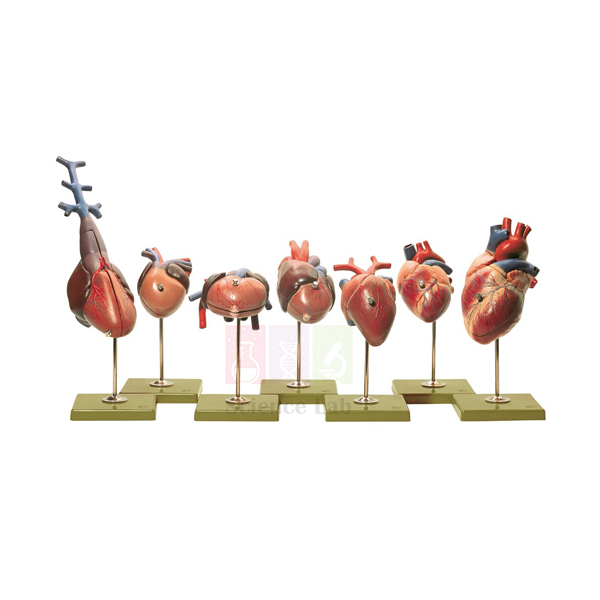
Vertebrate Hearts Model Set
Seven two-part heart models, each life size or slightly enlarged, let a class compare 2-, 3- and 4-chambered vertebrate hearts side by side. The direction of blood flow is marked on every model. Students open each heart to see its chambers and follow where oxygen-poor and oxygen-rich blood stay apart or can mix.
Vertebrate heart set specifications
| Parameter | Detail |
|---|---|
| Contents | 7 individual heart models |
| Model construction | Each model in 2 parts, for identifying external and internal structures |
| Scale | Life size or slightly enlarged |
| Blood flow | Direction of flow marked on each model |
| Heart types compared | 2-, 3- and 4-chambered hearts |
| Study areas | Comparative anatomy, circulatory system, evolution |
| Product code | SLE/A/113 |
| Minimum order | 10 units |
| Dispatch | Usually within 7 to 10 working days |
| Manufacturer | ScienceLab (Jain Scientific Equipments Pvt Ltd), Ambala, India |
Circulation lessons with the seven hearts
Least to most divided. Lay the seven hearts in a row from the fewest chambers to the most. Students open each two-part model, count its chambers and follow the marked flow direction. In a table they record where oxygen-poor and oxygen-rich blood stay separate and where the two can meet.
Zooming in on the human heart. Once the trend is clear, groups move to the Human Heart Model to name the four chambers, the valves and the great vessels. The Human Cardiac Beat And Blood Circulation model then shows how the right side sends blood to the lungs while the left side supplies the body.
The heart in its place. Show the Hen Model, whose sectioned side shows a bird's heart among its other organs, and the Frog Dissection Model for the chest of an amphibian. Students find the heart in each animal and name the organs that lie beside it.
Frequently Asked Questions About the Vertebrate Hearts Model Set
What does single circulation mean for a two-chambered heart?
A two-chambered heart, as in fish, has one atrium and one ventricle and carries only oxygen-poor blood. It pumps blood to the gills to pick up oxygen, and from there the blood flows on to the body before it returns. Blood passes through the heart once per circuit, so its pressure is low by the time it reaches the organs.
Why can blood mix in a three-chambered heart?
In amphibians, two atria empty into one ventricle. The right atrium brings oxygen-poor blood from the body and the left atrium brings oxygen-rich blood from the lungs, so some mixing happens in the shared chamber. The spongy ventricle wall and a spiral valve in the outflow vessel keep the two streams partly apart.
How does a reptile heart differ from an amphibian heart?
Most reptiles have a partial wall, or septum, across the ventricle, so the two kinds of blood mix less than in amphibians. The gap in that wall also lets many reptiles send blood past the lungs while diving or resting. Crocodilians go a step further, with a fully divided ventricle.
Why do birds and mammals have a fully divided four-chambered heart?
A complete septum keeps the two kinds of blood fully apart and creates two circuits, one to the lungs and one to the body. The body circuit can then run at high pressure without harming the delicate lungs. That gives the steady oxygen supply active, warm-blooded animals need. Birds and mammals reached this layout independently.
What is the difference between an atrium and a ventricle?
An atrium is a receiving chamber with a thin wall that collects blood returning to the heart. A ventricle has a thicker, muscular wall and pumps blood out into the arteries. Valves between the chambers and at the exits let blood move one way only and stop it flowing back.
Why compare several heart models instead of studying just one?
Each model shows one version of the same basic plan, so only the full set makes the pattern visible. Side by side, the seven hearts show how far the chambers are divided in each group and how the marked flow paths separate. A single human heart model gives detail, but it cannot show this variation across animal groups.
Heart, circulation and comparative anatomy models
- Human Heart Model
- Human Cardiac Beat And Blood Circulation
- Human Circulatory System Model
- Vertebrate Brains Model Set
- Hen Model
- Frog Dissection Model
To request a quotation, send the product name, code and quantity through our contact page or add the item to your enquiry cart.
Last updated: 14 November 2026