- Physics Lab Equipment
- Chemistry Lab Equipment
- Biology Lab Equipment
- Anatomical Lab Equipment
- Learning
- Educational Lab Equipment
- Magnets
- Agriculture
- Astronomy
- Earth Science
- Geography Lab Equipment
- Safety Lab Coat
- Laboratory
- Meteorological
- Electricity & Magnetism
- Heat
- School Lab Equipment
- Lab Glassware
- Science Lab Equipment
- Tvet Lab Equipment
- Robotics Kits Supplier and Manufacturer for Schools India
- Math Lab Equipment Manufacturers and Exporters in Ambala
- STEM Kits Manufacturers and Supplier India
- Engineering Lab Equipment
- Hospital Lab Equipment
- Water, Sanitation & Hygiene Supplies
- Cold Chain, Solar & Communication Equipment
- Nutrition
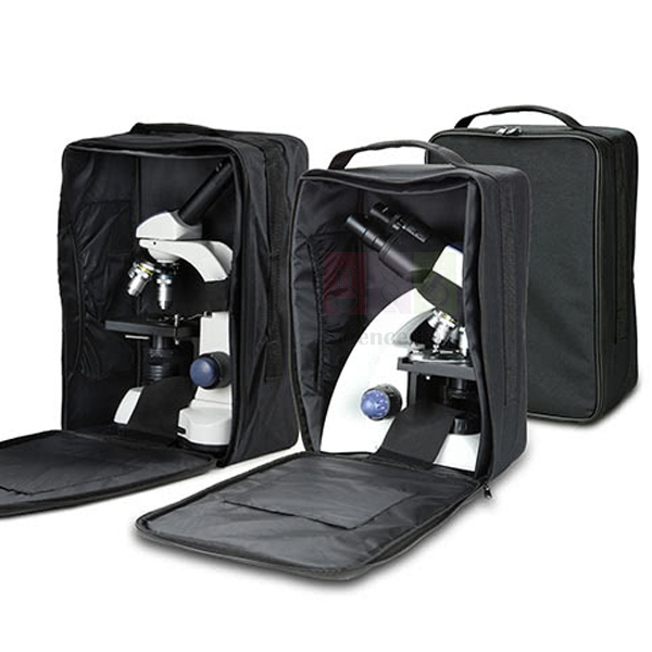
Carry Case for Microscopes
Carry Case for Microscopes (Product Code: SLE/M/126) is a nylon microscope bag used to keep a microscope completely dust free in storage and to carry it safely during field work. The microscope is placed inside easily and held in place with a few straps, and the bag measures 25 x 39 x 19 cm. It is supplied by Jain Scientific Equipments Pvt Ltd (ScienceLab), Ambala, India, for schools and field users.
Specifications
| Parameter | Detail |
|---|---|
| Type | Nylon microscope bag |
| Material | Nylon |
| Dimensions | 25 x 39 x 19 cm |
| Securing | A few straps hold the microscope safely inside the bag |
| Purpose | Dust-free storage; carrying a microscope on field work |
| Product code | SLE/M/126 |
| Supplied by | Jain Scientific Equipments Pvt Ltd (ScienceLab), Ambala, India |
What Is a Microscope Carry Case Used For?
Microscopes left uncovered in a school lab collect dust on the eyepieces, nosepiece and stage, and cleaning them before every class takes time. Keeping each microscope in its own nylon bag keeps it completely dust free between lessons, which saves time on cleaning and maintenance. For teachers running pond studies or ecology field trips, the same bag makes it easy to carry a microscope around all day.
Stand the microscope upright in the bag, fasten the straps around it and close the bag before lifting. Support the base of the bag when carrying, and compare the 25 x 39 x 19 cm size with your microscope's height, width and depth before ordering.
What to Specify When Ordering
- The microscope model the bag must hold, with its height, width and depth
- Number of bags, for example one per microscope
- Whether cleaning kits or lens tissue should be added to the order
Supplier
Jain Scientific Equipments Private Limited, trading as ScienceLab, supplies this nylon carry case from Ambala, Haryana, as an accessory for school and field microscopes. Schools, colleges, dealers and importers in India and international markets can order it with new microscopes or for instruments already in use. See the ScienceLab company profile for more.
Related Microscope Accessories
Storage alternatives, care items and portable microscopes:
- Microscope Case — alternative case for microscope storage
- Microscope Cleaning Kit — for cleaning a microscope after field use
- Lens Tissue — for wiping eyepieces and objectives on site
- Binocular Stereo Microscope, 40x — cordless stereo microscope for field work (confirm fit with us)
- Handheld Digital Microscope — portable option for outdoor observation
Browse more accessories in the Microscope category.
Frequently Asked Questions
What are the dimensions of the Carry Case for Microscopes?
The Carry Case for Microscopes measures 25 x 39 x 19 cm. Compare these dimensions with your microscope's height, width and depth, or send us the microscope model, and we will confirm whether it fits before you order.
What is the Carry Case for Microscopes made of?
The Carry Case for Microscopes is a nylon bag. The microscope is placed inside easily, and a few straps hold it safely in place, keeping it covered in storage and secure while it is carried.
Why store microscopes in a carry case?
Keeping each microscope in a Carry Case for Microscopes keeps it completely dust free between classes, which saves time on cleaning and maintenance of eyepieces, objectives and stage. It helps most in dusty rooms or where microscopes are stored for long periods.
Is the Carry Case for Microscopes suitable for field work?
Yes. The Carry Case for Microscopes is intended for users doing field work who need to carry a microscope around all day. Straps secure the microscope inside the bag, so it can be taken to a pond, garden or other field site.
Who supplies the Carry Case for Microscopes?
The Carry Case for Microscopes is supplied by Jain Scientific Equipments Pvt Ltd (ScienceLab), Ambala, Haryana, India, to schools, colleges, dealers and importers in India and international markets, alone or with microscopes.
What should a quotation request include?
For the Carry Case for Microscopes, send the number of bags, the microscope model or its dimensions, and the delivery location through the contact page or the enquiry cart, so we can confirm fit in the quotation.
Request a Quote
Ask for a quotation for the Carry Case for Microscopes through the contact page, or add it to the enquiry cart along with your microscopes.
Last updated: 24 September 2026

Prepared Microscope Slide Set – Health And Disease
Prepared Microscope Slide Set – Health And Disease (Product Code: SLE/PS/105) is a set of prepared glass microscope slides used to compare healthy and diseased human-body tissue in biology, health science and introductory pathology classes. It covers eight preparations with 2 slides of each, from lymph node and spleen to emphysema, thrombus, lung of smoking and atherosclerosis of the main and coronary arteries. It is supplied by Jain Scientific Equipments Pvt Ltd (ScienceLab), Ambala, India.
Specifications
| Parameter | Detail |
|---|---|
| Lymph node section | 2 slides |
| Spleen section | 2 slides |
| Spleen TS | 2 slides |
| Emphysema | 2 slides |
| Lung of smoking | 2 slides |
| Thrombus | 2 slides |
| Main artery atherosclerosis | 2 slides |
| Coronary artery atherosclerosis | 2 slides |
| Product code | SLE/PS/105 |
| Supplied by | Jain Scientific Equipments Pvt Ltd (ScienceLab), Ambala, India |
What Is the Prepared Microscope Slide Set – Health And Disease Used For?
The set brings disease topics from the textbook to the microscope. Lymph node and spleen slides show the organs where immune cells gather, giving students a reference for normal lymphoid tissue. The lung slides let them compare emphysema and the lung of a smoker, a strong visual link to lessons on smoking and gas exchange.
The cardiovascular slides cover a thrombus and atherosclerosis in both a main artery and a coronary artery, so a class can see how deposits narrow the vessel and discuss heart disease risk factors. With two copies of each preparation, one slide can stay on the demonstration microscope while the second passes round a small group. These prepared microscope slides also suit revision sessions before practical examinations.
What to Specify When Ordering
- Number of sets, bearing in mind each holds 2 slides per preparation
- Whether normal-tissue sets (such as circulatory or histology slides) should be quoted for comparison
- Storage and microscopes, if the lab needs them
Supplier
This slide set is supplied by Jain Scientific Equipments Private Limited, the company behind the ScienceLab name in Ambala, Haryana, for schools, colleges, training institutes and dealers in India and international markets. Institutions completing vendor forms can use the company's GSTIN 06AAECJ8618A1ZT and its Legal Entity Identifier 3358004HRD1JIVXKYX49. Visit our profile page to learn more.
Related Prepared Slides
Good companions for a health and disease unit:
- Prepared Microscope Slide Set – Circulatory System — healthy artery, vein and cardiac muscle for comparison with the diseased vessels
- Microslide Smoking & Health — annotated images of smoke-damaged airways and lungs
- Human Histology Slide Set of 25 — a wider set of normal human tissues
- Microscope Slide Case — protects the slides between classes
- Binocular Microscope, 1000x Mag LED — a microscope for detailed tissue work
More sets are listed under Prepared Slides.
Frequently Asked Questions
What slides are in the Prepared Microscope Slide Set – Health And Disease?
The Prepared Microscope Slide Set – Health And Disease contains lymph node section, spleen section, spleen TS, emphysema, thrombus, lung of smoking, main artery atherosclerosis and coronary artery atherosclerosis, with 2 slides of each preparation.
Why does the Health And Disease slide set include two of each slide?
The Prepared Microscope Slide Set – Health And Disease lists 2 slides per preparation, which lets a teacher keep one on a demonstration microscope while students examine the other, or keep a spare if a slide is damaged.
Is the Health And Disease slide set meant for diagnosis?
No. The Prepared Microscope Slide Set – Health And Disease is a teaching resource for showing students what diseased tissue looks like under a microscope. It is not intended for clinical or diagnostic use.
What microscope suits the Health And Disease slide set?
The Prepared Microscope Slide Set – Health And Disease can be examined with a standard compound microscope; higher magnification helps with cell detail in the lymph node, spleen and artery wall. We can quote microscopes on the same enquiry.
Who supplies the Prepared Microscope Slide Set – Health And Disease?
The Prepared Microscope Slide Set – Health And Disease is supplied by Jain Scientific Equipments Pvt Ltd (ScienceLab), Ambala, Haryana, India, which provides GSTIN and LEI details to institutions and importers on request.
What should an enquiry for the Health And Disease slide set include?
For the Prepared Microscope Slide Set – Health And Disease, send product code SLE/PS/105, the number of sets, any companion slide sets or microscopes, and the delivery location through our contact page.
Request a Quote
Request a quotation for the Prepared Microscope Slide Set – Health And Disease through our contact page, or add it to the enquiry cart with any other slide sets you are ordering.
Last updated: 24 September 2026

Prepared Microscope Slide Set – Exchange Surfaces and Epithelia
Prepared Microscope Slide Set – Exchange Surfaces and Epithelia (Product Code: SLE/PS/106) is a class set of prepared glass microscope slides used to study epithelial linings and the supporting tissues of gas exchange surfaces in biology practicals. It provides 10 slides each of ciliated epithelium, squamous epithelium, loose connective tissue, lung cartilage and hyaline cartilage, enough for groups to work on the same tissue at once. It is supplied by Jain Scientific Equipments Pvt Ltd (ScienceLab), Ambala, India.
Specifications
| Parameter | Detail |
|---|---|
| Ciliated epithelium | 10 slides |
| Squamous epithelium | 10 slides |
| Loose connective tissue | 10 slides |
| Lung cartilage | 10 slides |
| Hyaline cartilage | 10 slides |
| Slide type | Prepared microscope slides |
| Product code | SLE/PS/106 |
| Supplied by | Jain Scientific Equipments Pvt Ltd (ScienceLab), Ambala, India |
What Is the Prepared Microscope Slide Set – Exchange Surfaces and Epithelia Used For?
Exchange surfaces depend on thin linings and firm support, and this set lets students examine both. Squamous epithelium shows the flattened cells that keep diffusion distances short, while ciliated epithelium shows the hair-like cilia that sweep mucus along the airways. Loose connective tissue shows the packing layer that sits beneath such linings.
The lung cartilage and hyaline cartilage slides explain how the airways are held open, so students can relate structure to function when they draw and label the tissues. Because the set holds 10 of every preparation, a teacher can run one tissue per lesson with the whole class, or rotate the five preparations round stations. It is a practical microscope slide set for school and junior college biology labs preparing students for practical examinations.
What to Specify When Ordering
- Number of sets, based on 10 slides per preparation
- Whether linked sets such as Circulatory System or Connective Tissues should be quoted as well
- Microscopes or slide storage needed for the practical room
Supplier
ScienceLab, the trading name of Jain Scientific Equipments Private Limited in Ambala, Haryana, supplies this class set of epithelium and cartilage slides to schools, colleges and education distributors in India and international markets. We can combine it with other topic sets so a department receives one consolidated quotation. For company details, see our company profile.
Related Prepared Slides
Sets and equipment often ordered alongside:
- Prepared Microscope Slide Set – Connective Tissues — bone, adipose and areolar tissue to extend the tissue topic
- Prepared Microscope Slide Set – Circulatory System — the blood side of gas exchange
- Prepared Microscope Slide Set – Health And Disease — shows emphysema, where the exchange surface is damaged
- Monocular Biological Microscope 400x, LED — a student microscope for the practical
View the complete Prepared Slides range.
Frequently Asked Questions
What tissues are in the Exchange Surfaces and Epithelia slide set?
The Prepared Microscope Slide Set – Exchange Surfaces and Epithelia contains ciliated epithelium, squamous epithelium, loose connective tissue, lung cartilage and hyaline cartilage, with 10 slides of each preparation.
Is the Exchange Surfaces and Epithelia slide set enough for a whole class?
The Prepared Microscope Slide Set – Exchange Surfaces and Epithelia holds 10 slides of each tissue, so ten students or pairs can examine the same preparation together. Larger classes can order additional sets or rotate the slides between groups.
Why does the Exchange Surfaces and Epithelia set include cartilage?
The Prepared Microscope Slide Set – Exchange Surfaces and Epithelia includes lung and hyaline cartilage because cartilage supports the airways that carry air to the gas exchange surface, letting students connect lining tissues with the structures that keep air passages open.
What magnification is needed for the Exchange Surfaces and Epithelia slides?
The Prepared Microscope Slide Set – Exchange Surfaces and Epithelia is examined with a standard compound microscope; low power finds the tissue, and high power shows cell shape and cilia. We can quote suitable microscopes with the set.
Who supplies the Prepared Microscope Slide Set – Exchange Surfaces and Epithelia?
The Prepared Microscope Slide Set – Exchange Surfaces and Epithelia is supplied by Jain Scientific Equipments Pvt Ltd (ScienceLab), Ambala, Haryana, India, to schools, colleges and dealers in India and international markets.
Request a Quote
For a quotation on the Prepared Microscope Slide Set – Exchange Surfaces and Epithelia, contact us through our contact page or add it to the enquiry cart with the other sets your department needs.
Last updated: 24 September 2026

Prepared Microscope Slide Set – Circulatory System
Prepared Microscope Slide Set – Circulatory System (Product Code: SLE/PS/107) is a set of prepared glass microscope slides used for hands-on microscope practicals on blood, blood vessels, heart muscle and lymphatics. It contains 10 slides each of artery and vein, cardiac muscle, cardiac muscle (LS), human blood thin film and lymphatic (TS), so a full class can examine one preparation together. It is supplied by Jain Scientific Equipments Pvt Ltd (ScienceLab), Ambala, India.
Specifications
| Parameter | Detail |
|---|---|
| Artery and vein | 10 slides |
| Cardiac muscle | 10 slides |
| Cardiac muscle (LS) | 10 slides |
| Human blood thin film | 10 slides |
| Lymphatic (TS) | 10 slides |
| Product code | SLE/PS/107 |
| Supplied by | Jain Scientific Equipments Pvt Ltd (ScienceLab), Ambala, India |
What Is the Prepared Microscope Slide Set – Circulatory System Used For?
This set is designed for students to find structures themselves under the microscope. The artery and vein slide is the standard exercise for comparing wall thickness and lumen shape, and it usually leads to a labelled drawing. The blood thin film lets students tell red cells from the fewer white cells and practise counting fields of view.
Two cardiac muscle preparations, one of them a longitudinal section, give a class the chance to look for the striped, branching fibres of heart muscle, while the lymphatic transverse section completes the picture of how tissue fluid returns to the blood. With 10 copies of each slide, the preparations can rotate round the lab in one or two periods, which suits school biology lab equipment used for board practical preparation.
Supplier
The Circulatory System slide set comes from Jain Scientific Equipments Private Limited, trading as ScienceLab, of Ambala, Haryana. We supply prepared slide sets like this to schools, colleges and dealers across India and in international markets, and we can list them on the same quotation as microscopes and heart models. The company profile gives more background.
Related Prepared Slides
Resources that support a circulation practical:
- Microslide The Circulatory System — annotated images to preview before the practical
- Prepared Microscope Slide Set – Health And Disease — atherosclerosis and thrombus slides for comparison with healthy vessels
- Human Circulatory System Model — shows the route blood takes around the body
- Binocular Microscope, 1000x Mag LED — for close study of the blood film
- Immersion Oil, 25ml — for oil-immersion viewing of blood cells
Our Prepared Slides category lists the other sets.
Frequently Asked Questions
What does the Prepared Microscope Slide Set – Circulatory System contain?
The Prepared Microscope Slide Set – Circulatory System contains 10 slides each of five preparations: artery and vein, cardiac muscle, cardiac muscle (LS), human blood thin film, and lymphatic (TS).
How do the two cardiac muscle slides in the Circulatory System set differ?
In the Prepared Microscope Slide Set – Circulatory System, one cardiac muscle preparation is listed as LS, a longitudinal section, and the other simply as cardiac muscle. Ask us through the contact page if you need the orientation of the second one confirmed.
What magnification suits the blood film in the Circulatory System slide set?
The human blood thin film in the Prepared Microscope Slide Set – Circulatory System can be scanned at low and high power; many teachers use an oil-immersion objective with immersion oil to show white cell nuclei in detail.
How is the Circulatory System slide set different from the Microslide on the same topic?
The Prepared Microscope Slide Set – Circulatory System is made of glass slides for students to examine under a microscope, whereas Microslide The Circulatory System provides annotated photographic images that are looked at in a Microslide Viewer.
Who supplies the Prepared Microscope Slide Set – Circulatory System?
The Prepared Microscope Slide Set – Circulatory System is supplied by Jain Scientific Equipments Pvt Ltd (ScienceLab) in Ambala, Haryana, India, to schools, colleges and dealers in India and international markets.
What is needed for a quotation on the Circulatory System slide set?
For the Prepared Microscope Slide Set – Circulatory System, send code SLE/PS/107, the number of sets, any microscopes or immersion oil you want added, and the delivery location through our contact page.
Request a Quote
Get a quotation for the Prepared Microscope Slide Set – Circulatory System via our contact page, or add it to the enquiry cart together with microscopes and other slide sets.
Last updated: 24 September 2026

Prepared Microscope Slide Set – Transport In Plants
Prepared Microscope Slide Set – Transport In Plants (Product Code: SLE/PS/108) is a set of prepared glass microscope slides used to trace the path of water and food through roots, stems and leaves in plant biology practicals. It includes 5 slides each of eight preparations, from Vicia root hairs and sunflower roots to Helianthus, Zea and Tilia stems and the rolled leaf of marram grass. The set is supplied by Jain Scientific Equipments Pvt Ltd (ScienceLab), Ambala, India.
Specifications
| Parameter | Detail |
|---|---|
| Vicia (root apex) | 5 slides |
| Vicia young root hairs | 5 slides |
| Helianthus (sunflower), young and old root TS | 5 slides |
| Helianthus, stem, bundles TS | 5 slides |
| Helianthus, old stem, TS and LS | 5 slides |
| Zea stem TS and LS | 5 slides |
| Tilia old stem (RLS) | 5 slides |
| Ammophila (marram grass), leaf TS | 5 slides |
| Product code | SLE/PS/108 |
| Supplied by | Jain Scientific Equipments Pvt Ltd (ScienceLab), Ambala, India |
TS = transverse section; LS = longitudinal section; RLS = radial longitudinal section.
What Is the Prepared Microscope Slide Set – Transport In Plants Used For?
The slides follow water upward through the plant. Students begin at the Vicia root apex and young root hairs, where water enters, then examine young and old sunflower roots to locate the central xylem and phloem. Stem sections of Helianthus and Zea compare the ring of vascular bundles in a dicot with the scattered bundles of a monocot, and the TS and LS pairs show the same tissues cut two ways.
The old Helianthus and Tilia stems show how vascular tissue builds up as a stem ages, and the marram grass leaf, adapted to dry sand dunes, links transport to water loss and transpiration. Five copies of each slide allow small groups to share preparations, and teachers often run the set alongside a transpiration experiment so observation and measurement come together. It is a focused addition to any plant biology lab equipment list.
What to Specify When Ordering
- Number of sets for your class size (5 slides of each preparation per set)
- Whether single-copy reference sets, such as the Basic Plant Anatomy Slide Set, are also needed
- Any transpiration or root-pressure apparatus to include in the same quotation
Supplier
Jain Scientific Equipments Private Limited supplies the Transport In Plants slide set through its ScienceLab business in Ambala, Haryana. Schools, colleges and dealers in India and international markets order prepared slides from us together with botany apparatus, which keeps a department's purchasing on one quotation. Learn about the company on our profile page.
Related Prepared Slides
Items that complement a plant transport practical:
- Basic Plant Anatomy Slide Set — one-of-each reference slides of the same plants
- Kit for Demonstration of Transpiration in Plants — measures the water loss the leaf slides explain
- Kit for Demonstrating Root Pressure — shows water being pushed up from the roots
- Microslide The Root Of a Flowering Plant — annotated images of root zones and root hairs
- Monocular Biological Microscope 400x — a microscope for student groups
See more in Prepared Slides.
Frequently Asked Questions
Which slides are in the Prepared Microscope Slide Set – Transport In Plants?
The Prepared Microscope Slide Set – Transport In Plants has 5 slides each of Vicia root apex, Vicia young root hairs, sunflower young and old root TS, Helianthus stem bundles TS, Helianthus old stem TS and LS, Zea stem TS and LS, Tilia old stem RLS and marram grass leaf TS.
Does the Transport In Plants slide set show both monocot and dicot stems?
Yes. The Prepared Microscope Slide Set – Transport In Plants includes Zea (maize), a monocot, and Helianthus (sunflower) and Tilia (lime), both dicots, so students can compare how vascular bundles are arranged in each group.
Why is marram grass included in the Transport In Plants slide set?
The Prepared Microscope Slide Set – Transport In Plants includes an Ammophila (marram grass) leaf TS because this dune grass is a standard example of a leaf adapted to reduce water loss, connecting transport with transpiration.
How many students can use one Transport In Plants slide set?
Each Prepared Microscope Slide Set – Transport In Plants carries 5 slides of every preparation, so five students or groups can examine the same slide type at once. Order more sets for larger classes working simultaneously.
Who supplies the Prepared Microscope Slide Set – Transport In Plants?
The Prepared Microscope Slide Set – Transport In Plants is supplied by Jain Scientific Equipments Pvt Ltd (ScienceLab), Ambala, Haryana, India, for schools, colleges and dealers in India and international markets.
Request a Quote
Send an enquiry for the Prepared Microscope Slide Set – Transport In Plants on our contact page, or add it to the enquiry cart with any apparatus for the same practical.
Last updated: 24 September 2026

Prepared Microscope Slide Set – Connective Tissues
Prepared Microscope Slide Set – Connective Tissues (Product Code: SLE/PS/109) is a set of prepared glass microscope slides used to identify connective tissue, the three muscle types and related tissues in animal histology practicals. It supplies 5 slides each of eight preparations, including compact bone for Haversian canals and lamellae, adipose and areolar tissue, cardiac, striated and non-striated muscle, ciliated epithelium and spinal cord. It is supplied by Jain Scientific Equipments Pvt Ltd (ScienceLab), Ambala, India.
Specifications
| Parameter | Detail |
|---|---|
| Compact bone, for Haversian canals and lamellae | 5 slides |
| Adipose tissue, section | 5 slides |
| Areolar tissue, stained for fibres and cells E | 5 slides |
| Cardiac muscle showing the nuclei, striations and intercalated discs LS | 5 slides |
| Striated muscle, showing striations and nuclei LS | 5 slides |
| Non-striated (involuntary) muscle E | 5 slides |
| Pseudo-stratified ciliated epithelium TS | 5 slides |
| Spinal cord TS | 5 slides |
| Product code | SLE/PS/109 |
| Supplied by | Jain Scientific Equipments Pvt Ltd (ScienceLab), Ambala, India |
E = entire (whole mount); LS = longitudinal section; TS = transverse section.
What Is the Prepared Microscope Slide Set – Connective Tissues Used For?
The set supports the classic tissue identification practical. Students see how compact bone is organised into Haversian systems with rings of lamellae, how adipose tissue is packed with fat cells, and how areolar tissue, stained for fibres and cells, holds other tissues together. These three slides show why connective tissues look so different despite sharing a common role.
The muscle slides let a class compare the three types side by side: striated muscle with its striations and nuclei, cardiac muscle with intercalated discs, and non-striated (involuntary) muscle. Pseudo-stratified ciliated epithelium and a spinal cord section round out a survey of the main animal tissue types. With 5 slides of each, small groups can move through the preparations and record labelled drawings for their practical files.
Supplier
Jain Scientific Equipments Private Limited, which trades as ScienceLab in Ambala, Haryana, supplies the Connective Tissues slide set to schools, colleges, teaching laboratories and dealers in India and international markets. Buyers registering a new vendor can note the supplying company's Corporate Identification Number U29309HR2020PTC087597 and GSTIN 06AAECJ8618A1ZT. More on the company is on our profile page.
Related Prepared Slides
Other sets and tools used in histology practicals:
- Prepared Microscope Slide Set – Exchange Surfaces and Epithelia — more epithelium and cartilage slides
- Prepared Microscope Slide Set – Circulatory System — extra cardiac muscle and blood vessel slides
- Human Histology Slide Set of 25 — a broader single-copy reference set
- Binocular Biological Microscope — for examining tissue detail
Browse all Prepared Slides.
Frequently Asked Questions
What preparations are in the Prepared Microscope Slide Set – Connective Tissues?
The Prepared Microscope Slide Set – Connective Tissues contains compact bone, adipose tissue, areolar tissue, cardiac muscle LS, striated muscle LS, non-striated muscle, pseudo-stratified ciliated epithelium TS and spinal cord TS, with 5 slides of each.
Does the Connective Tissues slide set include muscle slides?
Yes. Besides bone, adipose and areolar tissue, the Prepared Microscope Slide Set – Connective Tissues includes cardiac, striated and non-striated muscle, plus ciliated epithelium and spinal cord, so all main animal tissue types can be compared.
What features does the compact bone slide in the Connective Tissues set show?
The compact bone slide in the Prepared Microscope Slide Set – Connective Tissues is prepared to show Haversian canals and lamellae, the concentric rings that make up the structural units of compact bone.
How many learners can share one Connective Tissues slide set?
One Prepared Microscope Slide Set – Connective Tissues provides 5 slides of each preparation, enough for five groups to view the same tissue together. Additional sets can be quoted for larger practical classes.
Who supplies the Prepared Microscope Slide Set – Connective Tissues?
The Prepared Microscope Slide Set – Connective Tissues is supplied by Jain Scientific Equipments Pvt Ltd (ScienceLab), Ambala, Haryana, India. CIN and GSTIN details are available to institutions that need them for vendor registration.
What should I send to get a quotation for the Connective Tissues slide set?
For the Prepared Microscope Slide Set – Connective Tissues, send code SLE/PS/109, the number of sets, any microscopes or companion slide sets, and your delivery city or country through our contact page.
Request a Quote
Request a quote for the Prepared Microscope Slide Set – Connective Tissues through our contact page, or add it to the enquiry cart along with other histology sets.
Last updated: 24 September 2026

Prepared Microscope Slide Set – Skin and Excretion
Prepared Microscope Slide Set – Skin and Excretion (Product Code: SLE/PS/110) is a group of ready-mounted microscope slides used to study skin structure and the organs of excretion in school and college biology practicals. It brings together two skin sections, a bladder-wall section and two kidney sections, so a class can follow how the body removes water, salts and wastes. These prepared microscope slides are supplied by Jain Scientific Equipments Pvt Ltd (ScienceLab), Ambala, India.
Key Details
| Parameter | Detail |
|---|---|
| Slide 1 | Skin, showing hair insertion and sebaceous glands, VS |
| Slide 2 | Skin, sweat glands, VS |
| Slide 3 | Transitional epithelium, bladder wall, TS |
| Slide 4 | Kidney, injected, thick section |
| Slide 5 | Kidney, section |
| Figure listed against each preparation | 10 (the listing does not say what this counts; the number of slides per preparation is confirmed in your quotation) |
| Label abbreviations | VS = vertical section; TS = transverse section |
| Product code | SLE/PS/110 |
| Supplied by | Jain Scientific Equipments Pvt Ltd (ScienceLab), Ambala, India |
What Is the Skin and Excretion Slide Set Used For?
Teachers use this set for practical lessons on excretion and temperature control. Students locate the hair follicle and its sebaceous gland on the first skin slide, then compare it with the coiled sweat glands on the second, which leads naturally into how sweating removes water and salts. The two kidney slides let the class pick out cortex and medulla, and the injected preparation, in which blood vessels are filled with a coloured medium, makes the kidney's rich blood supply easier to trace.
The bladder-wall slide shows transitional epithelium, the stretchable lining that allows the bladder to fill and empty. Begin every slide at low power to find the region of interest before moving to a higher objective. As part of school biology lab equipment, the set pairs well with a kidney model so students connect what they see under the lens with whole-organ anatomy.
What to Specify When Ordering
- The number of sets required, for example one per practical group or one for the teacher's demonstration.
- Confirmation of how many slides of each preparation you expect (the listing shows the figure 10 against each).
- Any microscopes, slide boxes or organ models to be quoted in the same enquiry.
Supplier
ScienceLab is the trading name of Jain Scientific Equipments Private Limited, based in Ambala, Haryana. The company supplies prepared slide sets like this one to schools, colleges, dealers and importers, and can combine them with microscopes and anatomy models so a department receives one consolidated quotation. More about the company is on the ScienceLab company profile.
Related Prepared Slides
These items are often ordered with the skin and excretion slides for the same practical sessions:
- Prepared Microscope Slide Set – Exchange Surfaces and Epithelia — other epithelium types to compare with the bladder lining.
- Human Kidney, Nephron, and Glomerulus Model Set — whole-organ and nephron view to match the kidney sections.
- Human Skin Section Model — enlarged 3D view of the glands seen on the skin slides.
- Monocular Biological Microscope 400x, LED — a classroom microscope for viewing the set.
- Slide Box — keeps the slides flat and protected between lessons.
See the full range in Prepared Slides.
Frequently Asked Questions
What slides are in the Prepared Microscope Slide Set – Skin and Excretion?
The Prepared Microscope Slide Set – Skin and Excretion lists five preparations: skin showing hair insertion and sebaceous glands (VS), skin sweat glands (VS), transitional epithelium of the bladder wall (TS), an injected thick kidney section and a plain kidney section.
How many slides of each preparation are supplied?
Our listing for the Skin and Excretion slide set shows the figure 10 beside each preparation but does not state what it counts. We therefore confirm the exact number of slides per preparation in your quotation; please ask through the contact page before you order.
What do VS and TS mean on the slide labels?
On the Skin and Excretion slide labels, VS means vertical section, cut at right angles to the skin surface, and TS means transverse section, cut straight across the organ. Knowing the plane of the cut helps students interpret the layers and glands they see.
What microscope is needed to view these slides?
A standard compound microscope with low- and high-power objectives is suitable for the Skin and Excretion prepared slides. Start at low power to find the gland or kidney region, then change to a higher objective; a school microscope can be quoted in the same enquiry.
How should the skin and excretion slides be stored?
Store the Skin and Excretion prepared slides flat in a slide box or tray, away from heat and direct sunlight, and hold them by the edges. Wipe fingerprints off gently with lens tissue and never let the objective lens press on the coverslip.
Who supplies the Skin and Excretion slide set?
Jain Scientific Equipments Pvt Ltd (ScienceLab), Ambala, India supplies the Prepared Microscope Slide Set – Skin and Excretion to schools, colleges, dealers and importers, handling quotations and orders for customers in India and international markets.
What information does a quotation request need?
To quote the Prepared Microscope Slide Set – Skin and Excretion, send product code SLE/PS/110, the number of sets, your delivery location and any microscopes or storage boxes you want added. The contact page and the enquiry cart both reach our sales team.
Request a Quote
Send your requirement for this slide set through the ScienceLab contact page, or add it to the enquiry cart with other prepared slides and microscopes for one combined quotation.
Last updated: 24 September 2026

Prepared Microscope Slide Set – Photosynthesis and Adaptations
Prepared Microscope Slide Set – Photosynthesis and Adaptations (Product Code: SLE/PS/111) is a collection of eight prepared plant slides used to show how leaves, stomata, pollen and root hairs are built for their jobs in secondary and college botany classes. Its mix of ordinary leaf sections with marram grass and fig preparations lets students compare typical leaves with plants adapted to harsher conditions. The set is supplied by Jain Scientific Equipments Pvt Ltd (ScienceLab), Ambala, India.
Key Details
| Parameter | Detail |
|---|---|
| Leaf sections | Dicot leaf, TS; Ligustrum leaf, TS; Prunus (cherry) leaf, TS |
| Stomata preparations | Monocot epidermis, for stomata, E; Ficus (fig), sunken stomata |
| Adaptation preparation | Ammophila (marram grass) |
| Reproduction and roots | Pine, mature pollen grain; Vicia, young root hairs |
| Preparations listed | 8 |
| Figure listed against each preparation | 5 (meaning not stated in the listing; slides per preparation confirmed at quotation) |
| Label abbreviations | TS = transverse section; E = entire (whole mount) |
| Product code | SLE/PS/111 |
| Supplied by | Jain Scientific Equipments Pvt Ltd (ScienceLab), Ambala, India |
What Is the Photosynthesis and Adaptations Slide Set Used For?
The three leaf sections are the core of a photosynthesis lesson: students identify the upper epidermis, the tightly packed palisade layer, the looser spongy tissue and the veins, then explain how each helps capture light and exchange gases. The monocot epidermis preparation shows stomata and guard cells from above, so learners can count pores or sketch their shape.
The remaining slides move the lesson towards adaptation. Marram grass is a sand-dune plant, and the fig preparation shows sunken stomata, both useful for discussing how plants limit water loss. Pine pollen introduces wind pollination, while the Vicia root-hair slide shows how roots increase their surface for absorbing water. Used with a photosynthesis experiment kit, the set links structure seen under the microscope with results measured at the bench, making it a practical piece of botany lab equipment.
What to Specify When Ordering
- How many sets you need for your practical groups.
- The number of slides of each preparation you expect, since the listing shows the figure 5 without explaining it.
- Whether photosynthesis apparatus or a microscope should be quoted alongside.
Supplier
Jain Scientific Equipments Private Limited, known to customers as ScienceLab, supplies this botany slide set from Ambala, Haryana. Botany departments, schools and resellers can place it on the same order as practical apparatus and other slide collections, which keeps paperwork and delivery simple. Company background is available on the ScienceLab profile page.
Related Prepared Slides
Items that support the same plant-biology practicals:
- Photosynthesis Apparatus — bench experiment to pair with the leaf sections.
- Prepared Microscope Slide Set – Transport In Plants — continues the topic into xylem and phloem.
- Microslide Photosynthesis — a viewer-based lesson on the same theme.
- Basic Plant Anatomy Slide Set — stems and roots to complete plant structure.
- Binocular Microscope 400x, LED Illumination — comfortable two-eye viewing of leaf tissues.
Browse all slide sets under Prepared Slides.
Frequently Asked Questions
Which plant preparations are in the Photosynthesis and Adaptations set?
The Prepared Microscope Slide Set – Photosynthesis and Adaptations lists eight preparations: dicot leaf TS, monocot epidermis for stomata, Ligustrum leaf TS, marram grass, mature pine pollen grain, fig sunken stomata, cherry leaf TS and Vicia young root hairs.
How many slides of each preparation does the set contain?
The Photosynthesis and Adaptations slide set listing shows the figure 5 beside each preparation without saying what it counts. We confirm the number of slides per preparation in writing with your quotation, so please raise this through the contact page when you enquire.
What does the letter E mean on some slide labels?
In the Photosynthesis and Adaptations set, E marks an entire or whole-mount preparation, such as the monocot epidermis peeled and mounted flat, whereas TS marks a thin transverse section cut across a leaf. Whole mounts show surface features like stomata in plan view.
Which slides illustrate adaptation to dry conditions?
In the Photosynthesis and Adaptations slide set, the marram grass and fig sunken-stomata preparations are the ones teachers usually use for water-saving adaptations. Comparing them with the ordinary dicot, privet and cherry leaf sections helps students explain why stomata position and leaf form differ.
Can these slides support a photosynthesis practical?
Yes, the Photosynthesis and Adaptations slide set supports the theory side of a photosynthesis practical by showing leaf tissues and stomata. It does not replace bench apparatus, so many schools order it together with photosynthesis apparatus to link observation with measured results.
Who supplies this slide set?
Jain Scientific Equipments Pvt Ltd (ScienceLab) of Ambala, India supplies the Prepared Microscope Slide Set – Photosynthesis and Adaptations to schools, colleges, laboratories and trade buyers in India and international markets, with quotations issued on request.
Request a Quote
For a quotation on code SLE/PS/111, write to us via the contact page or place the set in the enquiry cart with any apparatus or microscopes you need.
Last updated: 24 September 2026