- Physics Lab Equipment
- Chemistry Lab Equipment
- Biology Lab Equipment
- Anatomical Lab Equipment
- Learning
- Educational Lab Equipment
- Magnets
- Agriculture
- Astronomy
- Earth Science
- Geography Lab Equipment
- Safety Lab Coat
- Laboratory
- Meteorological
- Electricity & Magnetism
- Heat
- School Lab Equipment
- Lab Glassware
- Science Lab Equipment
- Tvet Lab Equipment
- Robotics Kits Supplier and Manufacturer for Schools India
- Math Lab Equipment Manufacturers and Exporters in Ambala
- STEM Kits Manufacturers and Supplier India
- Engineering Lab Equipment
- Hospital Lab Equipment
- Water, Sanitation & Hygiene Supplies
- Cold Chain, Solar & Communication Equipment
- Nutrition
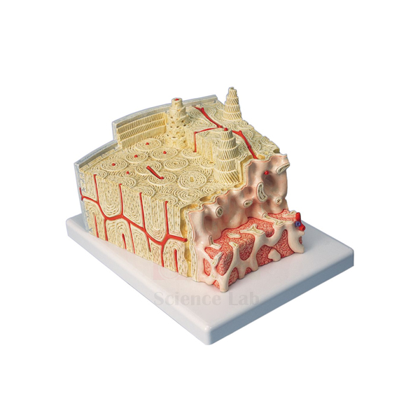
3B Bone Microanatomy Model
3B Bone Microanatomy Model (Product Code: SLE/A/89) enlarges a section of lamellar bone to about 80x life size so that the microscopic architecture of compact bone can be studied without a microscope. The block is cut in both cross and longitudinal section through all layers, down to the bone marrow, with Haversian canals, endosteum, cortical substance and osteocytes clearly visible. It is used in histology, physiology, orthopaedics and senior biology teaching, and is supplied by Jain Scientific Equipments Pvt Ltd (ScienceLab), Ambala, India.
Specifications
| Parameter | Detail |
|---|---|
| Scale | About 80x life size |
| Mounting | Mounted on a base |
| Tissue shown | Section of lamellar bone |
| Sections | Cross and longitudinal, through all layers including bone marrow |
| Visible structures | Haversian canals, endosteum, cortical substance, osteocytes |
Typical Use
Students often find it difficult to turn a flat slide image of compact bone into a mental picture of cylinders of lamellae running along the bone. Because this model shows the same tissue in two planes at once, a teacher can point to a Haversian canal in cross section and then follow it lengthwise in the longitudinal face, explaining how osteocytes sit between the lamellae and how the endosteum lines the inner surface next to the marrow.
It works best alongside the microscope: show the model first, then let learners find the same features on prepared microscope slides of bone. In physiology lessons it also supports discussion of bone remodelling and blood supply, while orthopaedic and physiotherapy students use it to understand how fractures heal at tissue level.
Supplier
This model is supplied by Jain Scientific Equipments Private Limited, which trades as ScienceLab from Ambala, Haryana, and sources selected teaching models to sit alongside its own laboratory products. For more about the company, see our company profile.
Related Human Models
Items that support a complete lesson on bone tissue and structure:
- Microslide Animal Tissue Set 2 Muscle, Bone, Connective — real bone sections to compare with the enlarged model
- Binocular Biological Microscope — for viewing bone slides after the model demonstration
- Human Bone Structures Model — shows bone at the level of the whole bone rather than the tissue
- Human Femur Bone Model — a long bone to relate the microanatomy to gross anatomy
- Skeletal Muscle Fiber Model — another enlarged tissue model for the musculoskeletal unit
Browse our anatomy models category for more microanatomy and organ models.
Frequently Asked Questions
How much is the bone enlarged?
About 80x life size, enough to make individual osteocytes and canals visible from across a classroom.
Which structures can students identify?
Haversian canals, endosteum, cortical substance, osteocytes and the bone marrow, shown in both cross and longitudinal section.
Is this model made by ScienceLab?
No. It is a branded model that we supply; our team can advise on availability and delivery for your order.
What are the dimensions and weight?
These are not given in the product details. Please ask through the contact page if you need them for storage or shipping.
How should it be cleaned?
Use a soft brush to remove dust from the textured surfaces and a slightly damp cloth on the base. Avoid solvents.
Can it be included in a bulk order for a medical college?
Yes. It can be quoted together with our own models for institutions in India and international markets; send your full list and delivery location.
Request a Quote
For availability and pricing, contact us on the contact page or place the model in your enquiry cart with the slides and microscope you need.
Last updated: 24 September 2026