- Physics Lab Equipment
- Chemistry Lab Equipment
- Biology Lab Equipment
- Anatomical Lab Equipment
- Learning
- Educational Lab Equipment
- Magnets
- Agriculture
- Astronomy
- Earth Science
- Geography Lab Equipment
- Safety Lab Coat
- Laboratory
- Meteorological
- Electricity & Magnetism
- Heat
- School Lab Equipment
- Lab Glassware
- Science Lab Equipment
- Tvet Lab Equipment
- Robotics Kits Supplier and Manufacturer for Schools India
- Math Lab Equipment Manufacturers and Exporters in Ambala
- STEM Kits Manufacturers and Supplier India
- Engineering Lab Equipment
- Hospital Lab Equipment
- Water, Sanitation & Hygiene Supplies
- Cold Chain, Solar & Communication Equipment
- Nutrition
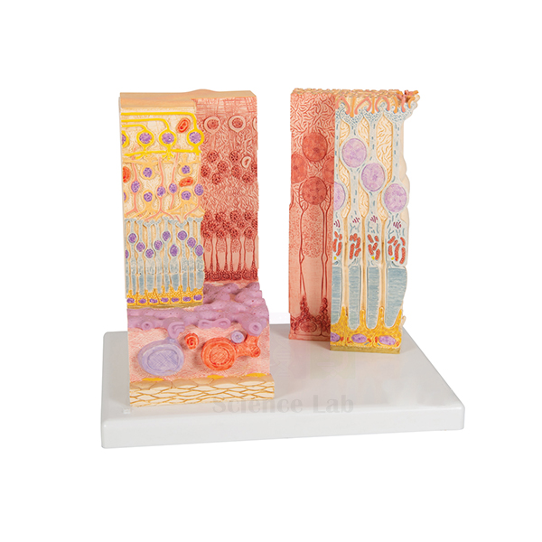
Eye Microanatomy Model
Eye Microanatomy Model (Product Code: SLE/A/80) brings the microscopic layers of the human retina up to a size a whole class can examine, presenting two highly magnified views side by side on one convenient base. It helps students connect what they read about rods, cones and neural layers with a three-dimensional form, and suits higher secondary biology, optometry, nursing and physiology teaching. The model is made by Jain Scientific Equipments Pvt Ltd (ScienceLab), Ambala, India.
Specifications
| Parameter | Detail |
|---|---|
| Subject | Microanatomy of the human retina |
| Views | Two highly magnified views on 1 base |
| Left view | Complete anatomical structure of the retina |
| Right view | Sectional enlargement of the retina |
| Size | 25 x 23 x 18.5 cm |
Typical Use
The retina is hard to picture from a textbook diagram, and this model fills that gap. A lecturer can start with the left-hand view to explain the overall organisation of the retina, then move to the sectional enlargement on the right to trace how light passes through the layers before reaching the photoreceptors. It works well at the start of a vision physiology unit, before students look at stained sections under the microscope, and as a revision aid when discussing conditions that affect the back of the eye.
Because both views share a single base, the model is easy to carry between classrooms and to place on a demonstration desk. Institutions building a set of eye model anatomy teaching aids in India often combine it with a whole-eyeball model, so learners can move from gross structure to the cellular level in one lesson.
Manufacturer
ScienceLab is the trading name of Jain Scientific Equipments Private Limited, which designs and supplies biology models and laboratory apparatus from Ambala in Haryana, India. Details of the company, its facilities and product lines are on our company profile.
Related Human Models
Items that support the same vision and eye-structure lessons:
- Human Eye Model — the whole eyeball, to set the retina in context
- Human Eye in Orbit Model — shows the eye, muscles and optic nerve within the bony orbit
- Cataract Eye Model with 4 Forms of Cataract — links normal eye anatomy to a common clinical condition
- Microslide Basic Human Histology — prepared human tissue slides for the microscope part of a histology practical
More eye, ear and organ models are listed in our anatomy models category.
Frequently Asked Questions
What part of the eye does this model show?
Only the retina. The left view shows its complete anatomical structure and the right view is a sectional enlargement, both highly magnified.
Is this a whole-eyeball model?
No. It is a microanatomy model of the retina. For the full eyeball, pair it with a separate eye model such as the Human Eye Model listed above.
What are the dimensions?
25 x 23 x 18.5 cm, with both views mounted on one base, so it fits easily on a teacher’s desk or display shelf.
What is the exact magnification?
The listing describes the views as highly magnified but does not state a factor. If you need the figure for a tender, ask us through the contact page.
Does it come with a labelled key?
A key is not mentioned in the product details. Please check with our team whether identification material can be provided with your order.
How do I keep the model in good condition?
Dust it with a soft brush, wipe with a slightly damp cloth when needed, and keep it out of direct sunlight to protect the painted layers.
What should an institutional quotation request include?
Mention the product code SLE/A/80, the quantity, the delivery address and any paperwork your purchase department needs, and we will prepare the quote.
Request a Quote
Send your requirement through our contact page, or place the model in the enquiry cart together with the other eye models you are considering.
Last updated: 24 September 2026